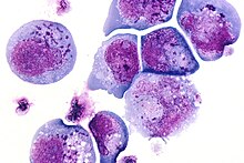
I-cell

Fritz Heinrich Jakob Lewy, a German-American neurologist, first identified and described inclusions in the brain cells of patients with Parkinson's disease and published his findings in the Lewandowsky's Handbook of Neurology in 1912.[2] I-cells also called inclusion cells are abnormal fibroblasts having a large number of dark inclusions in the cytoplasm of the cell (mainly in the central area). They are metabolically inactive structures of a cell and are not enclosed by a membrane.[3] The inclusions are of various fats, proteins, carbohydrates, pigments, excretory products, crystals,[4] and other insolubles. They are found in the cytoplasm of a cell in both prokaryotes and eukaryotes.[5] They are seen in Mucolipidosis II,[6] and Mucolipidosis III, also called inclusion-cell or I-cell disease where lysosomal enzyme transport and storage is affected.[7]


References
[edit]- ^ USA, Yale Rosen from (2011-08-04), The crystalline inclusions that may be found within the giant cells in sarcoidosis and other granulomatous disorders consist mainly of calcium oxalate and well as some calcium cargbonate. In this image there are multiple Schaumann bodies closely associated with crystalline inclusions. In many cases the crystals appear to serve as a nidus around which Schaumann bodies are formed. H&E stain., retrieved 2021-12-10
- ^ Engelhardt, Eliasz (October 2017). "Lafora and Trétiakoff: the naming of the inclusion bodies discovered by Lewy". Arquivos de Neuro-Psiquiatria. 75 (10): 751–753. doi:10.1590/0004-282X20170116. ISSN 0004-282X. PMID 29166468.
- ^ Lakna (2018-11-03). "What is the Difference Between Cell Organelles and Cell Inclusions". Pediaa.Com. Retrieved 2021-12-09.
- ^ Leliaert, Frederik; Coppejans, Eric (March 2004). "Crystalline cell inclusions: a new diagnostic character in the Cladophorophyceae (Chlorophyta)". Phycologia. 43 (2): 189–203. doi:10.2216/i0031-8884-43-2-189.1. ISSN 0031-8884. S2CID 86155723.
- ^ "inclusion body | biology | Britannica". britannica.com. Retrieved 2021-12-09.
- ^ al.], consultants Daniel Albert ... [et (2012). Dorland's illustrated medical dictionary (32nd ed.). Philadelphia, PA: Saunders/Elsevier. pp. 319 and 928. ISBN 978-1-4160-6257-8.
- ^ "Mucolipidoses Fact Sheet | National Institute of Neurological Disorders and Stroke". ninds.nih.gov. Retrieved 2021-12-09.
- ^ photographer, Unknown (October 1986), Deutsch: Lichtmikroskopische Aufnahme von infizierten T-Lymphozyten mit typischen sogenannten Einschlusskörperchen (HE-Färbung). Einschlüsse bei einer HHV6-Infektion (dunkelblaue Punkte)., retrieved 2021-12-10
- ^ Rajan, Suraj (2012-05-12), English: Photomicrograph of a region of substantia nigra in a Parkinson's patient showing Lewy bodies and Lewy neurites. It shows a 60-times magnification of the alpha synuclein intraneuronal inclusions aggregated to form Lewy bodies. Stains used: mouse monoclonal alpha-synuclein antibody; counterstained with Mayer's haematoxylin., retrieved 2021-12-10
