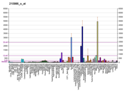
TPM1
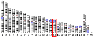
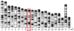
Tropomyosin alpha-1 chain is a protein that in humans is encoded by the TPM1 gene.[5] This gene is a member of the tropomyosin (Tm) family of highly conserved, widely distributed actin-binding proteins involved in the contractile system of striated and smooth muscles and the cytoskeleton of non-muscle cells.
Structure
[edit]Tm is a 32.7 kDa protein composed of 284 amino acids.[6] Tm is a flexible protein homodimer or heterodimer composed of two alpha-helical chains, which adopt a bent coiled coil conformation to wrap around the seven actin molecules in a functional unit of muscle.[7] It is polymerized end to end along the two grooves of actin filaments and provides stability to the filaments. Human striated muscles express protein from the TPM1 (α-Tm), TPM2 (β-Tm) and TPM3 (γ-Tm) genes, with α-Tm being the predominant isoform in striated muscle. In human cardiac muscle the ratio of α-Tm to β-Tm is roughly 5:1.[8]
Function
[edit]Tm functions in association with the troponin complex to regulate the calcium-dependent interaction of actin and myosin during muscle contraction. Tm molecules are arranged head-to-tail along the actin thin filament, and are a key component in cooperative activation of muscle. A three state model has been proposed by McKillop and Geeves,[9] which describes the positions of Tm during a cardiac cycle. The blocked (B) state occurs in diastole when intracellular calcium is low and Tm blocks the myosin binding site on actin. The closed (C) state is when Tm is positioned on the inner groove of actin; in this state myosin is in a "cocked" position where heads are weakly bound and not generating force. The myosin binding (M) state is when Tm is further displaced from actin by myosin crossbridges that are strongly-bound and actively generating force. In addition to actin, Tm binds troponin T (TnT). TnT tethers the region of head-to-tail overlap of subsequent Tm molecules to actin.
Clinical Significance
[edit]Mutations in TPM1 have been associated with hypertrophic cardiomyopathy (HCM), dilated cardiomyopathy and left ventricular noncompaction cardiomyopathy (LVNC). HCM mutations tend to cluster around the N-terminal region and a primary actin binding region known as period 5.[10]
References
[edit]- ^ a b c GRCh38: Ensembl release 89: ENSG00000140416 – Ensembl, May 2017
- ^ a b c GRCm38: Ensembl release 89: ENSMUSG00000032366 – Ensembl, May 2017
- ^ "Human PubMed Reference:". National Center for Biotechnology Information, U.S. National Library of Medicine.
- ^ "Mouse PubMed Reference:". National Center for Biotechnology Information, U.S. National Library of Medicine.
- ^ Mogensen J, Kruse TA, Borglum AD (Jun 1999). "Refined localization of the human alpha-tropomyosin gene (TPM1) by genetic mapping". Cytogenet Cell Genet. 84 (1–2): 35–6. doi:10.1159/000015207. PMID 10343096. S2CID 84901339.
- ^ "Protein Information". Archived from the original on September 24, 2015. Retrieved 25 May 2023.
{{cite web}}: CS1 maint: unfit URL (link) - ^ Brown, J. H.; Kim, K. H.; Jun, G; Greenfield, N. J.; Dominguez, R; Volkmann, N; Hitchcock-Degregori, S. E.; Cohen, C (2001). "Deciphering the design of the tropomyosin molecule". Proceedings of the National Academy of Sciences. 98 (15): 8496–501. Bibcode:2001PNAS...98.8496B. doi:10.1073/pnas.131219198. PMC 37464. PMID 11438684.
- ^ Yin, Z; Ren, J; Guo, W (2015). "Sarcomeric protein isoform transitions in cardiac muscle: A journey to heart failure". Biochimica et Biophysica Acta (BBA) - Molecular Basis of Disease. 1852 (1): 47–52. doi:10.1016/j.bbadis.2014.11.003. PMC 4268308. PMID 25446994.
- ^ McKillop, D. F.; Geeves, M. A. (1993). "Regulation of the interaction between actin and myosin subfragment 1: Evidence for three states of the thin filament". Biophysical Journal. 65 (2): 693–701. Bibcode:1993BpJ....65..693M. doi:10.1016/S0006-3495(93)81110-X. PMC 1225772. PMID 8218897.
- ^ Tardiff, J. C. (2011). "Thin filament mutations: Developing an integrative approach to a complex disorder". Circulation Research. 108 (6): 765–82. doi:10.1161/CIRCRESAHA.110.224170. PMC 3075069. PMID 21415410.
Further reading
[edit]- Lees-Miller JP, Helfman DM (1992). "The molecular basis for tropomyosin isoform diversity". BioEssays. 13 (9): 429–37. doi:10.1002/bies.950130902. PMID 1796905. S2CID 7958541.
- Balvay L, Fiszman MY (1995). "[Analysis of the diversity of tropomyosin isoforms]". C. R. Séances Soc. Biol. Fil. 188 (5–6): 527–40. PMID 7780795.
- Gunning P, Weinberger R, Jeffrey P (1997). "Actin and tropomyosin isoforms in morphogenesis". Anat. Embryol. 195 (4): 311–5. doi:10.1007/s004290050050. PMID 9108196. S2CID 9692297.
- Perry SV (2002). "Vertebrate tropomyosin: distribution, properties and function". J. Muscle Res. Cell. Motil. 22 (1): 5–49. doi:10.1023/A:1010303732441. PMID 11563548. S2CID 12346005.
- Perry SV (2004). "What is the role of tropomyosin in the regulation of muscle contraction?". J. Muscle Res. Cell. Motil. 24 (8): 593–6. doi:10.1023/B:JURE.0000009811.95652.68. PMID 14870975. S2CID 32621707.
- Jääskeläinen P, Miettinen R, Kärkkäinen P, et al. (2004). "Genetics of hypertrophic cardiomyopathy in eastern Finland: few founder mutations with benign or intermediary phenotypes". Ann. Med. 36 (1): 23–32. doi:10.1080/07853890310017161. PMID 15000344. S2CID 29985750.
- Mak A, Smillie LB, Bárány M (1978). "Specific phosphorylation at serine-283 of alpha tropomyosin from frog skeletal and rabbit skeletal and cardiac muscle". Proc. Natl. Acad. Sci. U.S.A. 75 (8): 3588–92. Bibcode:1978PNAS...75.3588M. doi:10.1073/pnas.75.8.3588. PMC 392830. PMID 278975.
- Höner B, Shoeman RL, Traub P (1992). "Degradation of cytoskeletal proteins by the human immunodeficiency virus type 1 protease". Cell Biol. Int. Rep. 16 (7): 603–12. doi:10.1016/S0309-1651(06)80002-0. PMID 1516138.
- Chevray PM, Nathans D (1992). "Protein interaction cloning in yeast: identification of mammalian proteins that react with the leucine zipper of Jun". Proc. Natl. Acad. Sci. U.S.A. 89 (13): 5789–93. Bibcode:1992PNAS...89.5789C. doi:10.1073/pnas.89.13.5789. PMC 402103. PMID 1631061.
- Shoeman RL, Kesselmier C, Mothes E, et al. (1991). "Non-viral cellular substrates for human immunodeficiency virus type 1 protease". FEBS Lett. 278 (2): 199–203. doi:10.1016/0014-5793(91)80116-K. PMID 1991513.
- Cho YJ, Liu J, Hitchcock-DeGregori SE (1990). "The amino terminus of muscle tropomyosin is a major determinant for function". J. Biol. Chem. 265 (1): 538–45. doi:10.1016/S0021-9258(19)40264-0. PMID 2136742.
- Colote S, Widada JS, Ferraz C, et al. (1988). "Evolution of tropomyosin functional domains: differential splicing and genomic constraints". J. Mol. Evol. 27 (3): 228–35. Bibcode:1988JMolE..27..228C. doi:10.1007/BF02100079. PMID 3138425. S2CID 24795087.
- Lin CS, Leavitt J (1988). "Cloning and characterization of a cDNA encoding transformation-sensitive tropomyosin isoform 3 from tumorigenic human fibroblasts". Mol. Cell. Biol. 8 (1): 160–8. doi:10.1128/mcb.8.1.160. PMC 363096. PMID 3336357.
- MacLeod AR, Gooding C (1988). "Human hTM alpha gene: expression in muscle and nonmuscle tissue". Mol. Cell. Biol. 8 (1): 433–40. doi:10.1128/mcb.8.1.433. PMC 363144. PMID 3336363.
- Mische SM, Manjula BN, Fischetti VA (1987). "Relation of streptococcal M protein with human and rabbit tropomyosin: the complete amino acid sequence of human cardiac alpha tropomyosin, a highly conserved contractile protein". Biochem. Biophys. Res. Commun. 142 (3): 813–8. doi:10.1016/0006-291X(87)91486-0. PMID 3548719.
- Heeley DH, Golosinska K, Smillie LB (1987). "The effects of troponin T fragments T1 and T2 on the binding of nonpolymerizable tropomyosin to F-actin in the presence and absence of troponin I and troponin C." J. Biol. Chem. 262 (21): 9971–8. doi:10.1016/S0021-9258(18)61061-0. PMID 3611073.
- Brown HR, Schachat FH (1985). "Renaturation of skeletal muscle tropomyosin: implications for in vivo assembly". Proc. Natl. Acad. Sci. U.S.A. 82 (8): 2359–63. Bibcode:1985PNAS...82.2359B. doi:10.1073/pnas.82.8.2359. PMC 397557. PMID 3857586.
- Tanokura M, Ohtsuki I (1984). "Interactions among chymotryptic troponin T subfragments, tropomyosin, troponin I and troponin C.". J. Biochem. 95 (5): 1417–21. doi:10.1093/oxfordjournals.jbchem.a134749. PMID 6746613.
- Pearlstone JR, Smillie LB (1983). "Effects of troponin-I plus-C on the binding of troponin-T and its fragments to alpha-tropomyosin. Ca2+ sensitivity and cooperativity". J. Biol. Chem. 258 (4): 2534–42. doi:10.1016/S0021-9258(18)32959-4. PMID 6822572.
External links
[edit]- Mass spectrometry characterization of human TPM1 at COPaKB [1]
- GeneReviews/NIH/NCBI/UW entry on Familial Hypertrophic Cardiomyopathy Overview
- Overview of all the structural information available in the PDB for UniProt: P09493 (Tropomyosin alpha-1 chain) at the PDBe-KB.
- ^ Zong, N. C.; Li, H; Li, H; Lam, M. P.; Jimenez, R. C.; Kim, C. S.; Deng, N; Kim, A. K.; Choi, J. H.; Zelaya, I; Liem, D; Meyer, D; Odeberg, J; Fang, C; Lu, H. J.; Xu, T; Weiss, J; Duan, H; Uhlen, M; Yates Jr, 3rd; Apweiler, R; Ge, J; Hermjakob, H; Ping, P (2013). "Integration of cardiac proteome biology and medicine by a specialized knowledgebase". Circulation Research. 113 (9): 1043–53. doi:10.1161/CIRCRESAHA.113.301151. PMC 4076475. PMID 23965338.
{{cite journal}}: CS1 maint: numeric names: authors list (link)

