Tumor necrosis factor superfamily
| Tumor necrosis factor superfamily | |||||||||
|---|---|---|---|---|---|---|---|---|---|
 Trimeric structure of TNF alpha, produced by Mus musculus, based on PDB structure 2TNF (1.4 Å Resolution). Different colors represent different monomers. Baeyens, KJ et al. (1999).[1] Figure rendered using FirstGlance Jmol. | |||||||||
| Identifiers | |||||||||
| Symbol | TNF | ||||||||
| Pfam | PF00229 | ||||||||
| InterPro | IPR006052 | ||||||||
| PROSITE | PDOC00224 | ||||||||
| SCOP2 | 1tnf / SCOPe / SUPFAM | ||||||||
| OPM superfamily | 292 | ||||||||
| OPM protein | 2hew | ||||||||
| Membranome | 80 | ||||||||
| |||||||||
| TNF | |||||||||
|---|---|---|---|---|---|---|---|---|---|
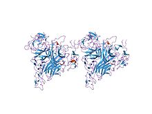
 crystal structure of trail-sdr5 | |||||||||
| Identifiers | |||||||||
| Symbol | TNF | ||||||||
| Pfam | PF00229 | ||||||||
| Pfam clan | CL0100 | ||||||||
| InterPro | IPR006052 | ||||||||
| PROSITE | PDOC00561 | ||||||||
| SCOP2 | 1tnr / SCOPe / SUPFAM | ||||||||
| |||||||||
The tumor necrosis factor (TNF) superfamily is a protein superfamily of type II transmembrane proteins containing TNF homology domain and forming trimers. Members of this superfamily can be released from the cell membrane by extracellular proteolytic cleavage and function as a cytokine. These proteins are expressed predominantly by immune cells and they regulate diverse cell functions, including immune response and inflammation, but also proliferation, differentiation, apoptosis and embryogenesis.[2][3]
The superfamily contains 19 members that bind to 29 members of TNF receptor superfamily.[4] An occurrence of orthologs in invertebrates hints at ancient origin of this superfamily in evolution.[2]
The PROSITE pattern of this superfamily is located in a beta sheet in the central section of the protein that is conserved across all members.
Members
[edit]There are 19 family members, numerically classified as TNFSF#, where # denotes the member number, sometimes followed by a letter.[4][2]
| TNFSF# | Name | Synonyms | Gene | Function |
|---|---|---|---|---|
| 1 | Lymphotoxin alpha | TNFβ, TNFSF1B | LTA | Induction of inflammation and antiviral response, development of secondary lymphoid organs, role in tumorigenesis |
| 2 | Tumor necrosis factor | TNFα, Dif, Necrosin, TNFSF1A, ... | TNF | Regulation of immune cells, induction of fever, cachexia, inflammation and apoptosis, inhibition of tumorigenesis and viral replication and response to sepsis |
| 3 | Lymphotoxin beta | TNFγ | LTB | Induction of inflammation and antiviral response, development of secondary lymphoid organs, role in tumorigenesis |
| 4 | OX40 ligand | CD252, Gp34, CD134L | TNFSF4 | Activation of T cell immune response by T cell costimulation |
| 5 | CD40 ligand | CD154, TRAP, Gp39, T-BAM | CD40LG | Regulation of adaptive immune response by activating antigen-presenting cell |
| 6 | Fas ligand | CD178, APTL, CD95L | FASLG | Regulation of T cell homeostasis by induction of apoptosis |
| 7 | CD27 ligand | CD70 | CD70 | Regulation of B cell activation and T cell homeostasis |
| 8 | CD30 ligand | CD153 | TNFSF8 | Induction of apoptosis of T cells and B cells, prevention of autoimmunity |
| 9 | CD137 ligand | 4-1 BBL | TNFSF9 | |
| 10 | TNF-related apoptosis-inducing ligand | CD253, APO-2L | TNFSF10 | Inhibition of tumorigenesis, induction of apoptosis |
| 11 | Receptor activator of nuclear factor kappa-Β ligand | CD254, OPGL, TRANCE, ODF | TNFSF11 | Tissue growth (particularly bone regeneration and remodeling), dendritic cell maturation |
| 12 | TNF-related weak inducer of apoptosis | APO-3L, DR3L | TNFSF12 | Regulation of angiogenesis, induction of apoptosis |
| 13 | A proliferation-inducing ligand | CD256, TALL-2, TRDL1 | TNFSF13 | Regulation of B cell development and plasma cell survival |
| 13B | B-cell activating factor | CD257, BLyS, TALL-1, TNFSF20, ... | TNFSF13B | Stimulation of B cell proliferation and differentiation |
| 14 | LIGHT | CD258, HVEML | TNFSF14 | Stimulation of T cell proliferation, apoptosis regulation |
| 15 | Vascular endothelial growth inhibitor | TL1, TL-1A | TNFSF15 | Inhibition of angiogenesis |
| 18 | TNF superfamily member 18 | GITRL, AITRL, TL-6 | TNFSF18 | Regulation of T cell survival |
| 19 | Ectodysplasin A | ED1-A1, ED1-A2 | EDA | Development of ectodermal tissues |
References
[edit]- ^ Baeyens KJ, De Bondt HL, Raeymaekers A, Fiers W, De Ranter CJ (April 1999). "The structure of mouse tumour-necrosis factor at 1.4 A resolution: towards modulation of its selectivity and trimerization". Acta Crystallographica Section D. 55 (Pt 4): 772–8. Bibcode:1999AcCrD..55..772B. doi:10.1107/s0907444998018435. PMID 10089307.
- ^ a b c The evolution of the immune system: conservation and diversification. Amsterdam: Elsevier. 2016. ISBN 978-0-12-801975-7. OCLC 950694824.
- ^ Abbas AK, Lichtman AH, Pillai S (May 2017). Cellular and molecular immunology (Ninth ed.). Philadelphia, PA. ISBN 978-0-323-47978-3. OCLC 973917896.
{{cite book}}: CS1 maint: location missing publisher (link) - ^ a b Aggarwal BB, Gupta SC, Kim JH (January 2012). "Historical perspectives on tumor necrosis factor and its superfamily: 25 years later, a golden journey". Blood. 119 (3): 651–65. doi:10.1182/blood-2011-04-325225. PMC 3265196. PMID 22053109.
External links
[edit]- Tumor+Necrosis+Factors at the U.S. National Library of Medicine Medical Subject Headings (MeSH)
- pex1 tumor necrosis factor gene
