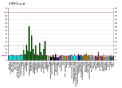
Microtubule-associated protein 2
Microtubule-associated protein 2 is a protein in humans that is encoded by the MAP2 gene.[5][6]
Function
[edit]This gene encodes a protein that belongs to the microtubule-associated protein family. The proteins of this family were originally isolated since they copurify with tubulin in polymerization experiments: tubulin in cell extracts can be made to polymerize to produce microtubules (MT) under the influence of heat and the addition of GTP, and the MT can then be collected by centrifugation. When this is done a series of microtubule associated proteins are collected along with the MT and can be detected by SDS-PAGE and other methods. Brain extracts are rich in several of these proteins, MAP2 being one of these. The single MAP2 gene produces four major transcripts producing four proteins, MAP2A, MAP2B, MAP2C and MAP2D. MAP2A and MAP2B are very high molecular weight proteins, with apparent molecular weight on SDS-PAGE about 250 kDa, while MAP2C and MAP2D are much lower molecular weight forms with apparent SDS-PAGE size about 70 kDa.[7] All forms of MAP2 share a common core sequence which includes MT binding domains, 18 amino acid sequences which are found in other MT associated proteins such as MAP Tau and MAP1B. The MAP2 isoforms are thought to be involved in MT assembly, which is an essential step in neuritogenesis. MAP2 serves to stabilize MT growth by crosslinking MT with intermediate filaments and other MTs. MAP2 isoforms are neuron-specific cytoskeletal proteins enriched in dendrites and perikarya, implicating a role in determining and stabilizing neuronal morphology during neuron development. As a result antibodies to MAP2 are widely used to identify neuronal cells and trace dendritic processes in experimental contexts.
Interactions
[edit]MAP2 has been shown to interact with Grb2,[8][9] NEFL[10] and MYO7A.[11] All MAP2 isoforms bind to microtubules.

References
[edit]- ^ a b c GRCh38: Ensembl release 89: ENSG00000078018 – Ensembl, May 2017
- ^ a b c GRCm38: Ensembl release 89: ENSMUSG00000015222 – Ensembl, May 2017
- ^ "Human PubMed Reference:". National Center for Biotechnology Information, U.S. National Library of Medicine.
- ^ "Mouse PubMed Reference:". National Center for Biotechnology Information, U.S. National Library of Medicine.
- ^ Neve RL, Harris P, Kosik KS, Kurnit DM, Donlon TA (May 1987). "Identification of cDNA clones for the human microtubule-associated protein tau and chromosomal localization of the genes for tau and microtubule-associated protein 2". Brain Res. 387 (3): 271–80. doi:10.1016/0169-328x(86)90033-1. PMID 3103857.
- ^ Kalcheva N, Albala J, O'Guin K, Rubino H, Garner C, Shafit-Zagardo B (December 1995). "Genomic structure of human microtubule-associated protein 2 (MAP-2) and characterization of additional MAP-2 isoforms". Proc Natl Acad Sci U S A. 92 (24): 10894–8. Bibcode:1995PNAS...9210894K. doi:10.1073/pnas.92.24.10894. PMC 40537. PMID 7479905.
- ^ "Entrez Gene: MAP2 microtubule-associated protein 2".
- ^ Lim RW, Halpain S (July 2000). "Regulated association of microtubule-associated protein 2 (MAP2) with Src and Grb2: evidence for MAP2 as a scaffolding protein". J. Biol. Chem. 275 (27): 20578–87. doi:10.1074/jbc.M001887200. PMID 10781592.
- ^ Zamora-Leon SP, Lee G, Davies P, Shafit-Zagardo B (October 2001). "Binding of Fyn to MAP-2c through an SH3 binding domain. Regulation of the interaction by ERK2". J. Biol. Chem. 276 (43): 39950–8. doi:10.1074/jbc.M107807200. PMID 11546790.
- ^ Frappier T, Stetzkowski-Marden F, Pradel LA (April 1991). "Interaction domains of neurofilament light chain and brain spectrin". Biochem. J. 275 (Pt 2): 521–7. doi:10.1042/bj2750521. PMC 1150082. PMID 1902666.
- ^ Todorov PT, Hardisty RE, Brown SD (March 2001). "Myosin VIIA is specifically associated with calmodulin and microtubule-associated protein-2B (MAP-2B)". Biochem. J. 354 (Pt 2): 267–74. doi:10.1042/0264-6021:3540267. PMC 1221652. PMID 11171103.
Further reading
[edit]- Roses AD, Einstein G, Gilbert J, Goedert M, Han SH, Huang D, Hulette C, Masliah E, Pericak-Vance MA, Saunders AM, Schmechel DE, Strittmatter WJ, Weisgraber KH, Xi PT (1996). "Morphological, biochemical, and genetic support for an apolipoprotein E effect on microtubular metabolism". Ann. N. Y. Acad. Sci. 777 (1): 146–57. Bibcode:1996NYASA.777..146R. doi:10.1111/j.1749-6632.1996.tb34413.x. PMID 8624078. S2CID 9145181.
- Snásel J, Pichová I (1997). "The cleavage of host cell proteins by HIV-1 protease". Folia Biol. (Praha). 42 (5): 227–30. doi:10.1007/BF02818986. PMID 8997639. S2CID 7617882.
- Shafit-Zagardo B, Kalcheva N (1999). "Making sense of the multiple MAP-2 transcripts and their role in the neuron". Mol. Neurobiol. 16 (2): 149–62. doi:10.1007/BF02740642. PMID 9588626. S2CID 2966442.
- Liu Y, Saad RS, Shen SS, Silverman JF (2003). "Diagnostic value of microtubule-associated protein-2 (MAP-2) for neuroendocrine neoplasms". Advances in Anatomic Pathology. 10 (2): 101–6. doi:10.1097/00125480-200303000-00005. PMID 12605092.
- Ainsztein AM, Purich DL (1992). "Cleavage of bovine brain microtubule-associated protein-2 by human immunodeficiency virus proteinase". J. Neurochem. 59 (3): 874–80. doi:10.1111/j.1471-4159.1992.tb08325.x. PMID 1494913. S2CID 40571317.
- Alberts MJ, Kandt RS, Pericak-Vance MA, Bebout J, Speer MC, Siddique TS, Yamaoka L, Hung WY, Gaskell PC, Roses AD (1991). "MspI RFLP for microtubule associated protein-2 (MAP2)". Nucleic Acids Res. 19 (4): 960. doi:10.1093/nar/19.4.960. PMC 333743. PMID 1708129.
- Wallin M, Deinum J, Goobar L, Danielson UH (1990). "Proteolytic cleavage of microtubule-associated proteins by retroviral proteinases". J. Gen. Virol. 71 (9): 1985–91. doi:10.1099/0022-1317-71-9-1985. PMID 2212989.
- Kosik KS, Orecchio LD, Bakalis S, Duffy L, Neve RL (1988). "Partial sequence of MAP2 in the region of a shared epitope with Alzheimer neurofibrillary tangles". J. Neurochem. 51 (2): 587–98. doi:10.1111/j.1471-4159.1988.tb01079.x. PMID 2455776. S2CID 31087371.
- Dammerman M, Yen SH, Shafit-Zagardo B (1990). "Sequence of a human MAP-2 region sharing epitopes with Alzheimer neurofibrillary tangles". J. Neurosci. Res. 24 (4): 487–95. doi:10.1002/jnr.490240405. PMID 2481044. S2CID 25209290.
- Obar RA, Dingus J, Bayley H, Vallee RB (1990). "The RII subunit of cAMP-dependent protein kinase binds to a common amino-terminal domain in microtubule-associated proteins 2A, 2B, and 2C". Neuron. 3 (5): 639–45. doi:10.1016/0896-6273(89)90274-2. PMID 2561973. S2CID 1329548.
- Rubino HM, Dammerman M, Shafit-Zagardo B, Erlichman J (1990). "Localization and characterization of the binding site for the regulatory subunit of type II cAMP-dependent protein kinase on MAP2". Neuron. 3 (5): 631–8. doi:10.1016/0896-6273(89)90273-0. PMID 2701845. S2CID 45499202.
- Herrmann H, Wiche G (1987). "Plectin and IFAP-300K are homologous proteins binding to microtubule-associated proteins 1 and 2 and to the 240-kilodalton subunit of spectrin". J. Biol. Chem. 262 (3): 1320–5. doi:10.1016/S0021-9258(19)75789-5. PMID 3027087.
- Garner CC, Tucker RP, Matus A (1989). "Selective localization of messenger RNA for cytoskeletal protein MAP2 in dendrites". Nature. 336 (6200): 674–7. doi:10.1038/336674a0. PMID 3200318. S2CID 4368119.
- Takahashi M, Tomizawa K, Sato K, Ohtake A, Omori A (1995). "A novel tau-tubulin kinase from bovine brain". FEBS Lett. 372 (1): 59–64. doi:10.1016/0014-5793(95)00955-9. PMID 7556643. S2CID 42295269.
- Kindler S, Garner CC (1995). "Four repeat MAP2 isoforms in human and rat brain". Brain Res. Mol. Brain Res. 26 (1–2): 218–24. doi:10.1016/0169-328X(94)90093-0. PMID 7854050.
- Albala JS, Kalcheva N, Shafit-Zagardo B (1994). "Characterization of the transcripts encoding two isoforms of human microtubule-associated protein-2 (MAP-2)". Gene. 136 (1–2): 377–8. doi:10.1016/0378-1119(93)90502-T. PMID 8294038.
- Illenberger S, Drewes G, Trinczek B, Biernat J, Meyer HE, Olmsted JB, Mandelkow EM, Mandelkow E (1996). "Phosphorylation of microtubule-associated proteins MAP2 and MAP4 by the protein kinase p110mark. Phosphorylation sites and regulation of microtubule dynamics". J. Biol. Chem. 271 (18): 10834–43. doi:10.1074/jbc.271.18.10834. PMID 8631898.
- Björkblom B, Ostman N, Hongisto V, Komarovski V, Filén JJ, Nyman TA, Kallunki T, Courtney MJ, Coffey ET (2005). "Constitutively active cytoplasmic c-Jun N-terminal kinase 1 is a dominant regulator of dendritic architecture: role of microtubule-associated protein 2 as an effector". J. Neurosci. 25 (27): 6350–61. doi:10.1523/JNEUROSCI.1517-05.2005. PMC 6725281. PMID 16000625.


