Трапезиометакарпал остеоартрит
| Трапезиометакарпал остеоартрит | |
|---|---|
| Другие имена | Карпометакарпал (CMC) остеоартрит (ОА) большого пальца, остеоартрит у основания большого пальца, базилярного (или базального) суставочного артрита, [ 1 ] Ризартроз [ 2 ] |
 | |
| Остеоартрит трапециометакарпального сустава | |
| Специальность | Пластическая хирургия |
Трапезиометакарпал остеоартрит (TMC OA) также известен как остеоартрит у основания эмпирического остеоартрита с эмпиасом, или в качестве ризартроза . [ 3 ] [ 1 ] [ 2 ] Этот сустав образуется трапециевой костью запястья и метакарпальной костью большого пальца. Это одно из суставов, где у большинства людей развивается остеоартрит с возрастом. [ 4 ] Остеоартрит-это возрастная потеря гладкой поверхности кости, где он движется к другой кости (хрящ сустава). [ 3 ] [ 5 ] В ответ на потерю хряща кости сгущаются на поверхности сустава, что приводит к субхондральному склерозу. Кроме того, костюмируются, называемые остеофитами (также известными как «костяные шпоры»), образуются на краях сустава. [ 6 ]
Основным симптомом является боль, особенно с захватом и защитой. [ 7 ] [ 8 ] Эта боль часто описывается как слабость, но истинная слабость не является частью этой болезни. Люди также могут отметить изменение формы большого пальца. [ 7 ] [ 8 ] Некоторые люди выбирают операцию, но большинство людей обнаруживают, что могут разместить трапециометакарпал -артрит. [ 9 ] [ 10 ] [ 11 ]
Признаки и симптомы
[ редактировать ]The symptom that brings people with TMC OA to the doctor is pain.[8] Pain is typically experienced with gripping and pinching. People experiencing pain may describe it as weakness.
There may be enlargement at the TMC joint.[8] This area may be tender, meaning it is painful when pressed. There may also be hyperextension of the metacarpophalangeal joint. The thumb metacarpal deviates towards the middle of the hand (adduction).[12] Also a grinding sound, known as crepitus, can be heard when the TMC joint is moved, more so when axial pressure is applied.[13]
Etiology and Epidemiology
[edit]TMC OA is an expected part of aging in men and women equally.[4] A population-based study of radiographic signs of pathophysiology in 3595 people assessed in a research-related comprehensive health examination found no association with physical workload.[9] A study of people seeing a hand specialist for symptoms unrelated to TMC OA demonstrated no relationship of radiographic TMC OA to hand activity.[14]
Studies that compare people presenting with TMC symptoms to people without symptoms are sometimes interpreted as indicating that activities can contribute to the development of TMC OA.[15] A more accurate conclusion may be that hand use is associated with seeking care for symptoms related to TMC OA. Ligamentous laxity is often associated with TMC OA, but this is based on rationale rather than experimental evidence.[16] Obesity may be related to TMC OA.[9]
Anatomy
[edit]The TMC joint is a synovial joint between the trapezium bone of the wrist and the metacarpal bone at the base of the thumb. This joint is a so-called saddle joint (articulatio sellaris), unlike the CMC joints of the other four fingers which are ellipsoid joints.[17] This means that the surfaces of the TMC joint are both concave and convex.
This shape provides the TMC joint a wide range of motion. Movements include:[18]
- Flexion
- Extension
- Abduction
- Adduction
- Opposition
- Reposition
- Circumduction
The TMC joint is stabilized by 16 ligaments.[19] Of these ligaments, the deep anterior oblique ligament, also known as the palmar beak ligament, is considered to be the most important stabilizing ligament.[20]

Diagnosis
[edit]TMC OA is diagnosed based on symptoms and signs.[8] Radiographs can confirm the diagnosis and the severity of TMC OA. Other diagnoses in this region include scaphotrapezial trapezoid arthritis and first dorsal compartment tendinopathy (De Quervain syndrome) although these are usually easy to distinguish.
Classification
[edit]TMC OA severity was classified by Eaton and Littler which can be simplified as follows:[21][22]
Stage 1:
- slight widening of the joint space
- < 1/3 subluxation of the joint (in any projection)
Stage 2:
- Osteophytes, < 2 mm in diameter, are present. (usually adjacent to the volar or dorsal facets of the trapezium)
Stage 3:
- Osteophytes, > 2 mm in diameter, are present (usually adjacent to the volar and dorsal facets of the trapezium)
- Slight joint space narrowing

Stage 4:
- Narrow joint space
- Concomitant scaphotrapezial arthritis
A simpler classification is no arthritis, some arthritis, and severe arthritis.[23] This simpler classification system omits the potentially contradictory details of the Eaton/Littler classification and keeps scaphotrapezial arthrosis separate.
Treatment
[edit]There are no treatments proved to slow or relieve TMC OA. In other words, there are no disease-modifying treatments. All treatments are symptom alleviating (palliative). Most surgery is reconstructive—it removes the TMC joint. Metacarpal osteotomy was proposed as a potentially disease modifying surgery for more limited arthrosis,[24] but there is no experimental support for this theory.[25]
There is limited and limited quality evidence regarding splints, corticosteroid injections, manual therapy and other palliative measures. Studies with adequate randomization, blinding, and independent assessment are lacking.
Arthrodesis fuses the TMC joint. It is uncommonly used.[26] Arthroplasty surgery for TMC OA removes part or all of the trapezium.[27] Surgery may also support the metacarpal by reconstructing a ligament using a tendon graft or weave. Surgery may also place something in the space where the trapeziometacarpal joint was, either a tendon wrapped up into a ball or a prosthesis.
The best available evidence suggests no difference in symptom alleviation with these variations of TMC arthroplasty.[28]

In one randomized trial comparing trapeziectomy alone with trapeziectomy with ligament reconstruction and trapeziectomy with ligament reconstruction and tendon interposition, patients evaluated 5 to 18 years after surgery had similar pain intensity, grip strength and key and tip pinch strengths after each procedure.[29] Trapeziectomy alone is associated with fewer complications than the other procedures.
Trapeziectomy
[edit]During trapeziectomy,[30] the trapezium bone is removed without any further surgical adjustments. The trapezium bone is removed through an approximately three centimeter long incision along the lateral side of the thumb. To preserve surrounding structures, the trapezium bone is removed "by splitting" it into pieces.
An empty gap is left by the trapeziectomy and the wound is closed with sutures. Despite this gap, no significant changes in function of the thumb are reported.[27] After the surgery, the thumb will be immobilized with a cast.
Trapeziectomy with tendon interposition
[edit]Some physicians still believe that it is better to fill the gap left by the trapeziectomy. They assume that filling the gap with a part of a tendon is preferable in terms of function, stability and position of the thumb. This is based on the assumption that interposition can help maintain the space between the metacarpal and the scaphoid, which will improve comfort and capability. Neither of these assumptions is supported by experimental evidence.
During trapeziectomy with TI, a longitudinal strip of the palmaris longus tendon is collected. [31] If this tendon is absent (which is the case in 13% of the population), half of the flexor carpi radialis tendon (FCR) can be used.
The tendon is then formed into a circular shape and placed in the gap, where it is stabilized by sutures.[12]
Trapeziectomy with ligament reconstruction
[edit]Another technique is used to reconstruct the volar beak ligament after trapeziectomy. The rationale is that ligament reconstruction(LR) helps maintain the gap between the metacarpal and the scaphoid, and that a larger gap is associated with greater comfort and capability.[32] Again these possibilities are not supported by experimental evidence.
During this procedure the anterior oblique ligament is reconstructed using the FCR tendon. There is a wide variety in techniques to perform this LR, but they all have a similar goal.
Trapeziectomy with LRTI
[edit]Some physicians believe that combining LR with TI will help maintain gap between the metacarpal and the scaphoid.[33] And that doing so will improve comfort and capability. Keep in mind that these aspects of the rationale are not supported by experimental evidence. The evidence suggests that all of these procedures have comparable long-term results.
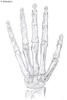
Arthrodesis
[edit]Arthrodesis of the TMC joint is a surgical procedure in which the trapezium bone and the metacarpal bone of the thumb are secured together. They are held together by K-wires or a plate and screws until the bone will heal.
Disadvantages include inability to flatten the hand.[27] Additionally, the stress on the CMC joint is now spread over the adjacent joints, those joints are more likely to develop osteoarthritis.[34]
Nevertheless, this procedure can be used in patients with stage II and III CMC OA as well as in young people with posttraumatic osteoarthritis.[27]
Joint replacement
[edit]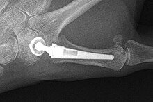
The joint can be replaced with artificial material. An artificial joint is also referred to as a prosthesis. Prostheses are more problematic at the trapeziometacarpal joint compared to joints like the knee or the hips.
[27]Prostheses come in many varieties, such as spacers or resurfacing prostheses.
It’s not clear within the current literature that a prosthesis has any advantage over trapeziectomy.[27]
Overall, joint replacements are related to long-term complications such as subluxation, fractures, synovitis (due to the material used) and nerve damaging.[35] In many cases revision surgery is needed to either remove or repair the prosthesis. Also note that usage of a joint replacement is heavy in costs.
The quality of the prostheses is improving and there is reason to believe this will have a positive effect on outcome in the years to follow.[27]

Metacarpal osteotomy
[edit]The aim of metacarpal osteotomy is to change the pressure distribution on the TMC joint. The hope is that this will slow the pace of development of osteoarthritis. There is no evidence that this procedure can modify the natural course of TMC OA. Osteotomy may be considered for people with mild arthritis.[24]
During osteotomy, the metacarpal is cut and a wedge shape bone fragment is removed to move the bone away from the hand.[36] Postoperative, the thumb of the patient is immobilized using a thumb-cast.
Possible complications are non-union of the bone, persistent pain related to unrecognized CMC or pantrapezial disease and radial sensory nerve injury.[24]
Complications
[edit]The most common complication after surgery is pain persisting in the thumb. Over long term, there is pain relief, but on short term, patients experience pain from the surgery itself. The main complaint is a burning sensation or hypersensitivity over the incision. Some patients develop a complex regional pain syndrome. This is a syndrome of chronic pain with changes of temperature and colour of the skin.
Other general complications include superficial radial nerve damage and postoperative wound infection.
After arthrodesis, non-union, in which fusion of the trapezium bone with the metacarpal bone fails, occurs in 8% to 21% of the cases.[27]
Subluxation of a prosthesis is a complication where the prosthesis is mobile and is partially dislocated. When the prosthesis is fully dislocated it is called a luxation. Both are painful and need revision surgery so the prosthesis can be repaired or removed.[37] When using a prosthesis over a longer period of time, there is a chance of breaking the prosthesis itself. This is due to mechanical wear.
Prostheses might also cause a reaction of the body against the artificial material they are made of, resulting in local inflammation.
Epidemiology
[edit]CMC OA is the most common form of OA affecting the hand.[38] Dahaghin et al. showed that about 15% of women and 7% of men between 50 and 60 years of age develop CMC OA of the thumb.[39] However, in about 65% of people older than 55 years, radiologic evidence of OA was present without any symptoms.[39] Armstrong et al. reported a prevalence of 33% in postmenopausal women, of which one-third was symptomatic, compared to 11% in men older than 55 years.[38] This shows CMC OA of the thumb is significantly more prevalent in women, especially in postmenopausal women, compared to men.
References
[edit]- ^ Jump up to: a b Patel, Tejas J.; Beredjiklian, Pedro K.; Matzon, Jonas L. (2012-12-16). "Trapeziometacarpal joint arthritis". Current Reviews in Musculoskeletal Medicine. 6 (1): 1–8. doi:10.1007/s12178-012-9147-6. ISSN 1935-973X. PMC 3702767. PMID 23242976.
- ^ Jump up to: a b TACCARDO, GIUSEPPE; DE VITIS, ROCCO; PARRONE, GIUSEPPE; MILANO, GIUSEPPE; FANFANI, FRANCESCO (2014-01-08). "Surgical treatment of trapeziometacarpal joint osteoarthritis". Joints. 1 (3): 138–144. ISSN 2512-9090. PMC 4295705. PMID 25606524.
- ^ Jump up to: a b Pelligrini VD (November 1991). "Osteoarthritis of the trapeziometacarpal joint: the pathophysiology of articular cartilage degeneration. II. Articular wear patterns in the osteoarthritic joint". J Hand Surg Am. 16 (6): 975–82. doi:10.1016/S0363-5023(10)80055-3. PMID 1748768.
- ^ Jump up to: a b Becker, Stéphanie J. E.; Briet, Jan Paul; Hageman, Michiel G. J. S.; Ring, David (December 2013). "Death, taxes, and trapeziometacarpal arthrosis". Clinical Orthopaedics and Related Research. 471 (12): 3738–3744. doi:10.1007/s11999-013-3243-9. ISSN 1528-1132. PMC 3825869. PMID 23959907.
- ^ Kihara H (April 1992). "[Anatomical study of the normal and degenerative articular surfaces on the first carpometacarpal joint]". Nippon Seikeigeka Gakkai Zasshi (in Japanese). 66 (4): 228–39. PMID 1593195.
- ^ North ER, Eaton RG (March 1983). "Degenerative joint disease of the trapezium: a comparative radiographic and anatomic study". J Hand Surg Am. 8 (2): 160–6. doi:10.1016/s0363-5023(83)80008-2. PMID 6833724.
- ^ Jump up to: a b Burton R. I. (1973). "Basal joint arthrosis of the thumb". Orthop. Clin. North Am. 4 (347): 331–8. doi:10.1016/S0030-5898(20)30797-5. PMID 4707436.
- ^ Jump up to: a b c d e Glickel SZ (May 2001). "Clinical assessment of the thumb trapeziometacarpal joint". Hand Clin. 17 (2): 185–95. doi:10.1016/S0749-0712(21)00239-0. PMID 11478041.
- ^ Jump up to: a b c Haara, Mikko M.; Heliövaara, Markku; Kröger, Heikki; Arokoski, Jari P. A.; Manninen, Pirjo; Kärkkäinen, Alpo; Knekt, Paul; Impivaara, Olli; Aromaa, Arpo (July 2004). "Osteoarthritis in the carpometacarpal joint of the thumb. Prevalence and associations with disability and mortality". The Journal of Bone and Joint Surgery. American Volume. 86 (7): 1452–1457. doi:10.2106/00004623-200407000-00013. ISSN 0021-9355. PMID 15252092.
- ^ Wilkens, Suzanne C.; Ring, David; Teunis, Teun; Lee, Sang-Gil P.; Chen, Neal C. (March 2019). "Decision Aid for Trapeziometacarpal Arthritis: A Randomized Controlled Trial". The Journal of Hand Surgery. 44 (3): 247.e1–247.e9. doi:10.1016/j.jhsa.2018.06.004. ISSN 1531-6564. PMID 30031600. S2CID 51709751.
- ^ Becker, Stéphanie J. E.; Teunis, Teun; Blauth, Johann; Kortlever, Joost T. P.; Dyer, George S. M.; Ring, David (March 2015). "Medical services and associated costs vary widely among surgeons treating patients with hand osteoarthritis". Clinical Orthopaedics and Related Research. 473 (3): 1111–1117. doi:10.1007/s11999-014-3912-3. ISSN 1528-1132. PMC 4317453. PMID 25171936.
- ^ Jump up to: a b Ghavami A, Oishi SN (May 2006). "Thumb trapeziometacarpal arthritis: treatment with ligament reconstruction tendon interposition arthroplasty". Plast. Reconstr. Surg. 117 (6): 116e–128e. doi:10.1097/01.prs.0000214652.31293.23. PMID 16651933. S2CID 6741548.
- ^ Carr M. M., Freiberg A. (1994). "Osteoarthritis of the thumb: Clinical aspects and management". Am. Fam. Physician. 50 (995): 995–1000. PMID 7942418.
- ^ Wilkens, Suzanne C.; Tarabochia, Matthew A.; Ring, David; Chen, Neal C. (May 2019). "Factors Associated With Radiographic Trapeziometacarpal Arthrosis in Patients Not Seeking Care for This Condition". Hand (New York, N.Y.). 14 (3): 364–370. doi:10.1177/1558944717732064. ISSN 1558-9447. PMC 6535938. PMID 28918660.
- ^ Fontana, Luc; Neel, Stéphanie; Claise, Jean-Marc; Ughetto, Sylvie; Catilina, Pierre (April 2007). "Osteoarthritis of the thumb carpometacarpal joint in women and occupational risk factors: a case-control study". The Journal of Hand Surgery. 32 (4): 459–465. doi:10.1016/j.jhsa.2007.01.014. ISSN 0363-5023. PMID 17398355.
- ^ Doerschuk SH, Hicks DG, Chinchilli VM, Pellegrini VD (May 1999). "Histopathology of the palmar beak ligament in trapeziometacarpal osteoarthritis". J Hand Surg Am. 24 (3): 496–504. doi:10.1053/jhsu.1999.0496. PMID 10357527.
- ^ Sobotta Anatomy, 14th ed., Elsevier, ISBN 9780702034831
- ^ Gray's Anatomy for Students, second edition, Elsevier, ISBN 9780443069529
- ^ Bettinger PC, Linscheid RL, Berger RA, Cooney WP, Kai-Nan A (1999). "An anatomic study of the stabilizing ligaments of the trapezium and trapeziometacarpal joint". J Hand Surg Am. 24 (4): 786–798. doi:10.1053/jhsu.1999.0786. PMID 10447171.
- ^ Pellegrini VD, Olcott CW, Hollenberg G (March 1993). "Contact patterns in the trapeziometacarpal joint: the role of the palmar beak ligament". J Hand Surg Am. 18 (2): 238–44. doi:10.1016/0363-5023(93)90354-6. PMID 8463587.
- ^ Томаино, MM Thumb Basal Count Arthritis. В DP Green et al. (Ред.), Оперативная операция Грина, 5 -е изд. Нью -Йорк: Черчилль Ливингстон, 2005. С. 461–485.
- ^ Eaton RG, Glickel SZ (ноябрь 1987 г.). «Трапезиометакарпал остеоартрит. Постановка в качестве обоснования лечения». Ручная клиника . 3 (4): 455–71. doi : 10.1016/s0749-0712 (21) 00761-7 . PMID 3693416 .
- ^ Содха, Самир; Кольцо, Дэвид; Зураковски, Дэвид; Юпитер, Джесси Б. (декабрь 2005 г.). «Распространенность остеоартроза сустава трапециометакарпала» . Журнал хирургии костей и суставов. Американский том . 87 (12): 2614–2618. doi : 10.2106/jbjs.e.00104 . ISSN 0021-9355 . PMID 16322609 .
- ^ Jump up to: а беременный в Atroshi I, Axelsson G, Nilsson EL (июнь 1998 г.). «Артропластика остеотомии в сравнении с сухожилиями при арпезиометакарпале артроза: 17 пациентов следовали за 1 год» . Acta Orthop Scand . 69 (3): 287–90. doi : 10.3109/17453679809000932 . PMID 9703405 .
- ^ Бачура, Абдо; Якиш, Эрик Дж.; Любан, Джон Д. (август 2018 г.). «Выживаемость и долгосрочные результаты остеотомии удлинения метакарпала с эмпиатром при симптоматической плотности карпометакарпала и раннего базального суставочного артрита» . Журнал хирургии рук . 43 (8): 772.e1–772.e7. doi : 10.1016/j.jhsa.2018.01.005 . ISSN 1531-6564 . PMID 29503049 . S2CID 3709534 .
- ^ Вольф, Дженнифер Мориатис; Деларонде, Стивен (январь 2012 г.). «Современные тенденции в неоперативном и оперативном лечении трапециометакарпального остеоартрита: обследование ручных хирургов США» . Журнал хирургии рук . 37 (1): 77–82. doi : 10.1016/j.jhsa.2011.10.010 . ISSN 1531-6564 . PMID 22119601 .
- ^ Jump up to: а беременный в дюймовый и фон глин час Вермейлен, Гус М.; Slijper, вред; Фейц, Рейнье; Hovius, Стивен ER; Moojen, Thybout M.; Selles, Ruud W. (2011). «Хирургическое лечение первичного большого пальца карпометакарпального остеоартрита: систематический обзор». Журнал хирургии рук . 36 (1): 157–169. doi : 10.1016/j.jhsa.2010.10.028 . ISSN 0363-5023 . PMID 21193136 .
- ^ Ваджон, Энн; Виникомб, Тоби; Карр, Эмма; Эдмундс, Ян; Ада, Луиза (2015-02-23). Ваджон, Энн (ред.). «Хирургия по поводу остеоартрита пальца (трапециометакарпал)» . Кокрановская база данных систематических обзоров . 2015 (2): CD004631. doi : 10.1002/14651858.cd004631.pub4 . ISSN 1469-493X . PMC 6464627 . PMID 25702783 .
- ^ Гангопадхьяй С., МакКенна Х, Берк Ф.Д., Дэвис Т.Р. (март 2012 г.). «Пяти-18 лет наблюдения за лечением трапециометакарпального остеоартрита: проспективное сравнение удаления, межпозиция сухожилий и реконструкции связок и сухожилия». J Hand Surg Am . 37 (3): 411–7. doi : 10.1016/j.jhsa.2011.11.027 . PMID 22305824 .
- ^ Жервис В.П. (1949). «Исключение трапеции для остеоартрита сустава трапециометакарпала». J Bone Suct Surg . 31b (4): 537–539. doi : 10.1302/0301-620x.31b4.537 . PMID 15397137 .
- ^ Вейльби А (1988). «Артропластика сухожилия первого карпо-мета-мета-карпального сустава». J Hand Surg Br . 13 (4): 421–425. doi : 10.1016/0266-7681 (88) 90171-4 . PMID 3249143 .
- ^ Blank J, Feldon P (1997). «Стабилизация метакарпофалангического суставов во время хирургии суставов карпометакарпала». Атлас Ханд Клини . 2 : 217–225.
- ^ Бертон Р.И., Пеллегрини В.Д. (май 1986 г.). «Хирургическое лечение базального сустава артрита большого пальца. Часть II. Реконструкция связки с артропластикой сухожилий». J Hand Surg Am . 11 (3): 324–32. doi : 10.1016/s0363-5023 (86) 80137-x . PMID 3711604 .
- ^ Mureau MA, Rademaker RP, Verhaar JA, Hovius SE (сентябрь 2001 г.). «Артропластика сухожилия в зависимости от артродеза для лечения трапециометакарпального артрита: ретроспективное сравнительное последующее исследование». J Hand Surg Am . 26 (5): 869–76. doi : 10.1053/JHSU.2001.26659 . PMID 11561240 .
- ^ Wajon A, Vinycomb T, Carr E, Edmunds I, Ada L (февраль 2015 г.). Ваджон А (ред.). «Хирургия по поводу остеоартрита пальца (трапециометакарпал)» . Кокрановская база данных Syst Rev. 2015 (2): CD004631. doi : 10.1002/14651858.cd004631.pub4 . PMC 6464627 . PMID 25702783 . (Втянут, см Два : 10.1002/14651858.cd004631.pub5 , PMID 28368089 )
- ^ Hobby JL, Lyall HA, Meggitt BF (май 1998). «Первая метакарпальная остеотомия при трапециометакарпале остеоартрит» . J Bone Suct Surg Br . 80 (3): 508–12. doi : 10.1302/0301-620x.80b3.8199 . PMID 9619947 .
- ^ Вейльби А., Сёндорф Дж (март 1978 г.). «Результаты после удаления силиконовых метакарпальных имплантатов трапеции». J Hand Surg Am . 3 (2): 154–6. doi : 10.1016/s0363-5023 (78) 80064-1 . PMID 632545 .
- ^ Jump up to: а беременный Армстронг А.Л., Хантер Дж.Б., Дэвис ТРК (1994). «Распространенность репаративного артрита основания большого пальца у женщин в постменопаузе». J Hand Surg . 19b (3): 340–341. doi : 10.1016/0266-7681 (94) 90085-X . PMID 8077824 . S2CID 26424006 .
- ^ Jump up to: а беременный Dahaghin S, Bierma-Zeinstra SM, Ginai AZ, Pols HA, Hazes JM, Koes BW (май 2005 г.). «Распространенность и схема рентгенографического остеоартрита рук и связь с болью и инвалидностью (исследование Роттердама)» . Энн. Рев. Диск . 64 (5): 682–7. doi : 10.1136/ard.2004.023564 . PMC 1755481 . PMID 15374852 .