Желток
| Желток | |
|---|---|
 Человеческий эмбрион 3,6 мм | |
 Human embryo from thirty-one to thirty-four days | |
| Details | |
| Carnegie stage | 5b |
| Days | 9 |
| Precursor | Endoderm |
| Identifiers | |
| Latin | vesicula umbilicalis; saccus vitellinus |
| MeSH | D015017 |
| TE | sac_by_E5.7.1.0.0.0.4 E5.7.1.0.0.0.4 |
| FMA | 87180 |
| Anatomical terminology | |
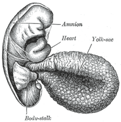
Желтый мешок представляет собой мембранный мешок, прикрепленный к эмбриону , образованный клетками слоя гипобласта биламинского эмбрионального диска . Это альтернативно называется пупочной пузырькой Terminologia Emryologica (TE), хотя желток гораздо более широко используется. У людей желточный мешок важен в раннем эмбриональном кровоснабжении, [ 1 ] и большая часть этого включена в изначальную кишку в течение четвертой недели эмбрионального развития . [ 2 ]
У людей
[ редактировать ]

Желтый мешок - первый элемент, увиденное в гестационном мешке во время беременности , [ 1 ] Обычно в 3 дня беременности .
Желток расположен на передней ( вентральной ) части эмбриона ; он выложен в экстра- эндодерме , [ 3 ] за пределами которого находится слой внеэмбриональной мезенхимы , полученный из эпибласта.
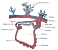
Blood is conveyed to the wall of the yolk sac by the primitive aorta and after circulating through a wide-meshed capillary plexus, is returned by the vitelline veins to the tubular heart of the embryo. This constitutes the vitelline circulation, which in humans serves as a location of haematopoiesis.[4][5] Before the placenta is formed and can take over, the yolk sac provides nutrition and gas exchange between the mother and the developing embryo.[6]
At the end of the fourth week, the yolk sac presents the appearance of a small pear-shaped opening (traditionally called the umbilical vesicle), into the digestive tube by a long narrow tube, the vitelline duct. Rarely, the yolk sac can be seen in the afterbirth as a small, somewhat oval-shaped body whose diameter varies from 1 mm to 5 mm; it is situated between the amnion and the chorion and may lie on or at a varying distance from the placenta. There is no clinical significance to a residual external yolk sac.
-
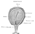
 Diagram showing earliest observed stage of human ovum.
1 - Amniotic cavity
2 - Yolk-sac
3 - Chorion -
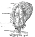
 Diagram illustrating early formation of allantois and differentiation of body-stalk.
1 Amniotic cavity
2 Body-stalk
3 Allantois
4 Yolk-sac
5 Chorion -
 Diagram showing later stage of allantoic development with commencing constriction of the yolk-sac.
1 Heart
2 Amniotic cavity
3 Embryo
4 Body-stalk
5 Placental villi
6 Allantois
7 Yolk-sac
8 Chorion -
 Diagram illustrating a later stage in the development of the umbilical cord.
1 Placental villi
2 Yolk-sac
3 Umbilical cord
4 Allantois
5 Heart
6 Digestive tube
7 Embryo
8 Amniotic cavity


As a rule the duct undergoes complete obliteration by the 20th week as most of the yolk sac is incorporated into the developing gastrointestinal tract, but in about two percent of cases its proximal part persists as a diverticulum from the small intestine, Meckel's diverticulum, which is situated about 60 cm proximal to the ileocecal valve, and may be attached by a fibrous cord to the abdominal wall at the umbilicus.
Sometimes a narrowing of the lumen of the ileum is seen opposite the site of attachment of the duct.
Histogenesis
[edit]The yolk sac starts forming during the second week of the embryonic development, at the same time as the shaping of the amniotic sac. The hypoblast starts proliferating laterally and descending. In the meantime Heuser's membrane, located on the opposite pole of the developing vesicle, starts its upward proliferation and meets the hypoblast.
Modifications
[edit]- Primary yolk sac: it is the vesicle which develops in the second week, its floor is represented by Heuser's membrane and its ceiling by the hypoblast. It is also known as the exocoelomic cavity.
- Secondary yolk sac: this structure is formed when the extraembryonic mesoderm separates to form the extraembryonic coelom; cells from the mesoderm pinch off an area of the yolk sac,[3] and what remains is the secondary yolk sac.
- The final yolk sac: during the fourth week of development, during organogenesis, part of the yolk sac is surrounded by endoderm and incorporated into the embryo as the gut. The remaining part of the yolk sac is the final yolk sac.
Additional images
[edit]-
 Surface view of embryo of Hylobates concolor (a gibbon).
 -
 Human embryo—length, 2 mm. Dorsal view, with the amnion laid open. X 30.
 -
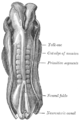 Dorsum of human embryo, 2.11 mm in length.
 -
 Section through the embryo.
 -
 Fetus of about eight weeks, enclosed in the amnion. Magnified a little over two diameters.
 -
 Модель человеческого эмбриона длиной 1,3 мм.
 -
 Раздел через яйцеклет
 -
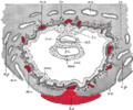
 Человеческий эмбрион около пятнадцати дней. Мозг и сердце представлены с правой стороны. Пищеварительная трубка и желток в срединной секции.


Смотрите также
[ редактировать ]Дальнейшее чтение
[ редактировать ]- Го, Иссак; и др. (18 августа 2023 г.). «Атлас клеток желточного мешка выявляет многоорганные функции во время раннего развития человека» . Наука . 381 (6659). doi : 10.1126/science.add7564 . PMC 7614978 .
Ссылки
[ редактировать ]- ^ Подпрыгнуть до: а беременный Лутфей, Карен; Фриз, Джереми (2005). «На пути к некоторым основаниям фундаментальной причинно -следственной связи: социально -экономический статус и здоровье в обычном посещении клиники для диабета» . Американский журнал социологии . 110 (5): 1326–1372. doi : 10.1086/428914 . ISSN 0002-9602 . JSTOR 10.1086/428914 . S2CID 17629087 .
- ^ Развивающийся человек: клинически ориентированная анатомия: глава 7
- ^ Подпрыгнуть до: а беременный Hafez, S. (2017-01-01), Huckle, William R. (ed.), "Chapter One - Comparative Placental Anatomy: Divergent Structures Serving a Common Purpose", Progress in Molecular Biology and Translational Science, Molecular Biology of Placental Development and Disease, 145, Academic Press: 1–28, doi:10.1016/bs.pmbts.2016.12.001, PMID 28110748, retrieved 2020-10-21
- ^ Мур, Кит; Персо, ТВН; Торчия, Марк (2013). Развивающийся человек . Филадельфия, Пенсильвания: Сондерс. ISBN 978-1-4377-2002-0 .
- ^ Blaas, Harm-Gerd K; Каррера, Хосе М. (2009-01-01), Wladimiroff, Juryy W; Eik-Nes, Sturla H (Eds.), «Глава 4-Исследование ранней беременности» , УЗИ в акушерстве и гинекологии , Эдинбург: Elsevier, стр. 57–78, DOI : 10.1016/B978-0-444-51829-3.00004. -0 , ISBN 978-0-444-51829-3 Получено 2020-10-21
- ^ Донован, Мэри Ф.; Bordoni, Bruno (2020), «Эмбриология, желток» , Statpearls , Остров Сокровища (FL): Patpearls Publishing, PMID 32310425 , получен 2020-09-11
| Базы данных управления авторитетом : национальный |
|---|