Подоцит
| Подоцит | |
|---|---|
 Подоциты, показанные зелеными, капсулы линии Боумена в почечной корпуску и обертывают вокруг капилляров в качестве основной части процесса фильтрации в почках | |
| Подробности | |
| Предшественник | Промежуточная мезодерма |
| Расположение | Боумена почек Капсула |
| Идентификаторы | |
| латинский | Podocytus |
| Сетка | D050199 |
| FMA | 70967 |
| Анатомические термины микроанатомии | |
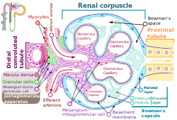
Подоциты представляют собой клетки в капсуле Боумена в почках которые обертываются вокруг капилляров клубочка . , Подоциты составляют эпителиальную подкладку капсулы Боумена, третий слой, через который происходит фильтрация крови. [ 1 ] Капсула Боумена фильтрует кровь , сохраняя большие молекулы , такие как белки, в то время как меньшие молекулы, такие как вода , соли и сахар, фильтрованы в качестве первого этапа образования мочи . Хотя различные визгуры имеют эпителиальные слои, название висцеральных эпителиальных клеток обычно относится конкретно к подоцитам, которые представляют собой специализированные эпителиальные клетки, которые находятся в висцеральном слое капсулы.
Подоциты имеют длинные первичные процессы, называемые трабекулами , которые образуют вторичные процессы, известные как цветоножки или процессы ноги (для которых клетки называются Podo - + -цит ). [ 2 ] Циллинки обернуты вокруг капилляров и оставляют прорезь между ними. Кровь фильтруется через эти прорезь, каждая из которых известна как щельфинальная щель , щель диафрагма или щели . [ 3 ] Несколько белков необходимы для того, чтобы цветоножки обернулись вокруг капилляров и функции. Когда дети рождаются с определенными дефектами в этих белках, таких как нефрин и CD2AP , их почки не могут функционировать. У людей есть различия в этих белках, и некоторые вариации могут предрасполагать их к почечной недостаточности в более позднем возрасте. Нефрин представляет собой белок, похожий на молнию , который образует щель диафрагму, с пространствами между зубами на молнии, достаточно большим, чтобы обеспечить сахар и воду, но слишком малы, чтобы пропускать белки. Дефекты нефрина ответственны за врожденную почечную недостаточность. CD2AP регулирует цитоскелет Podocyte и стабилизирует щель диафрагмы. [ 4 ] [ 5 ]
Структура
[ редактировать ]
Подоцит имеет сложную структуру. Его клеточный корпус имеет расширение основных или первичных процессов, которые образуют вторичные процессы в виде для ног подоцитов . процессов или цветоногих [ 6 ] Основные процессы удерживаются микротрубочками и промежуточными филаментами . Процессы ног имеют цитоскелет на основе актина. [ 6 ] Подоциты найдены в подкладке капсулы Боумена в нефронах почки. Процессы цветоножки или ног обертывают гломерулярные капилляры , чтобы сформировать щели фильтрации. [ 7 ] Плодовицы увеличивают площадь поверхности ячеек, обеспечивая эффективную ультрафильтрацию . [ 8 ]

Podocytes secrete and maintain the basement membrane.[3]
There are numerous coated vesicles and coated pits along the basolateral domain of the podocytes which indicate a high rate of vesicular traffic.
Podocytes possess a well-developed endoplasmic reticulum and a large Golgi apparatus, indicative of a high capacity for protein synthesis and post-translational modifications.
There is also growing evidence of a large number of multivesicular bodies and other lysosomal components seen in these cells, indicating a high endocytic activity.
Function
[edit]
A. The endothelial cells of the glomerulus; 1. pore (fenestra).
B. Glomerular basement membrane: 1. lamina rara interna 2. lamina densa 3. lamina rara externa
C. Podocytes: 1. enzymatic and structural protein 2. filtration slit 3. diaphragma
Podocytes have primary processes called trabeculae, which wrap around the glomerular capillaries.[2] The trabeculae in turn have secondary processes called pedicels or foot processes.[2] Pedicels interdigitate, thereby giving rise to thin gaps called filtration slits.[3] The slits are covered by slit diaphragms which are composed of a number of cell-surface proteins including nephrin, podocalyxin, and P-cadherin, which restrict the passage of large macromolecules such as serum albumin and gamma globulin and ensure that they remain in the bloodstream.[9] Proteins that are required for the correct function of the slit diaphragm include nephrin,[10] NEPH1, NEPH2,[11] podocin, CD2AP.[12] and FAT1.[13]

Small molecules such as water, glucose, and ionic salts are able to pass through the filtration slits and form an ultrafiltrate in the tubular fluid, which is further processed by the nephron to produce urine.
Podocytes are also involved in regulation of glomerular filtration rate (GFR). When podocytes contract, they cause closure of filtration slits. This decreases the GFR by reducing the surface area available for filtration.
Clinical significance
[edit]
A loss of the foot processes of the podocytes (i.e., podocyte effacement) is a hallmark of minimal change disease, which has therefore sometimes been called foot process disease.[15]
Disruption of the filtration slits or destruction of the podocytes can lead to massive proteinuria, where large amounts of protein are lost from the blood.
An example of this occurs in the congenital disorder Finnish-type nephrosis, which is characterised by neonatal proteinuria leading to end-stage kidney failure. This disease has been found to be caused by a mutation in the nephrin gene.
In 2002 Professor Moin Saleem at the University of Bristol made the first conditionally immortalised human podocyte cell line.[16][further explanation needed] This meant that podocytes could be grown and studied in the lab. Since then many discoveries have been made. Nephrotic syndrome occurs when there is a breakdown of the glomerular filtration barrier. The podocytes form one layer of the filtration barrier. Genetic mutations can cause podocyte dysfunction leading to an inability of the filtration barrier to restrict urinary protein loss. There are currently 53 genes known to play a role in genetic nephrotic syndrome.[17] In idiopathic nephrotic syndrome, there is no known genetic mutation. It is thought to be caused by a hitherto unknown circulating permeability factor.[18] Recent evidence suggests that the factor could be released by T-cells or B-cells,[19][20] podocyte cell lines can be treated with plasma from patients with nephrotic syndrome to understand the specific responses of the podocyte to the circulating factor. There is growing evidence that the circulating factor could be signalling to the podocyte via the PAR-1 receptor.[21][further explanation needed]
Presence of podocytes in urine has been proposed as an early diagnostic marker for preeclampsia.[22]
See also
[edit]- List of human cell types derived from the germ layers
- List of distinct cell types in the adult human body
References
[edit]- ^ "Podocyte" at Dorland's Medical Dictionary
- ^ Jump up to: a b c Ovalle WK, Nahirney PC (28 February 2013). Netter's Essential Histology E-Book. Elsevier Health Sciences. ISBN 978-1-4557-0307-4. Retrieved 2 June 2020.
- ^ Jump up to: a b c Lote CJ (2012). "Glomerular Filtration". Principles of Renal Physiology (5th ed.). New York: Springer Science+Business Media. p. 34. doi:10.1007/978-1-4614-3785-7_3. ISBN 978-1-4614-3784-0.
- ^ Wickelgren I (October 1999). "First components found for new kidney filter". Science. 286 (5438): 225–226. doi:10.1126/science.286.5438.225. PMID 10577188. S2CID 43237744.
- ^ Löwik MM, Groenen PJ, Levtchenko EN, Monnens LA, van den Heuvel LP (November 2009). "Molecular genetic analysis of podocyte genes in focal segmental glomerulosclerosis--a review". European Journal of Pediatrics. 168 (11): 1291–1304. doi:10.1007/s00431-009-1017-x. PMC 2745545. PMID 19562370.
- ^ Jump up to: a b Reiser J, Altintas MM (2016). "Podocytes". F1000Res. 5: 114. doi:10.12688/f1000research.7255.1. PMC 4755401. PMID 26918173.
- ^ Histology image:22401lba from Vaughan, Deborah (2002). A Learning System in Histology: CD-ROM and Guide. Oxford University Press. ISBN 978-0195151732.
- ^ Nosek TM. "Epithelium; Cell Types". Essentials of Human Physiology. Archived from the original on 24 March 2016.
- ^ Jarad G, Miner JH (May 2009). "Update on the glomerular filtration barrier". Current Opinion in Nephrology and Hypertension. 18 (3): 226–232. doi:10.1097/mnh.0b013e3283296044. PMC 2895306. PMID 19374010.
- ^ Wartiovaara J, Ofverstedt LG, Khoshnoodi J, Zhang J, Mäkelä E, Sandin S, et al. (November 2004). "Nephrin strands contribute to a porous slit diaphragm scaffold as revealed by electron tomography". The Journal of Clinical Investigation. 114 (10): 1475–1483. doi:10.1172/JCI22562. PMC 525744. PMID 15545998.
- ^ Neumann-Haefelin E, Kramer-Zucker A, Slanchev K, Hartleben B, Noutsou F, Martin K, et al. (June 2010). "A model organism approach: defining the role of Neph proteins as regulators of neuron and kidney morphogenesis". Human Molecular Genetics. 19 (12): 2347–2359. doi:10.1093/hmg/ddq108. PMID 20233749.
- ^ Fukasawa H, Bornheimer S, Kudlicka K, Farquhar MG (July 2009). "Slit diaphragms contain tight junction proteins". Journal of the American Society of Nephrology. 20 (7): 1491–1503. doi:10.1681/ASN.2008101117. PMC 2709684. PMID 19478094.
- ^ Ciani L, Patel A, Allen ND, ffrench-Constant C (May 2003). "Mice lacking the giant protocadherin mFAT1 exhibit renal slit junction abnormalities and a partially penetrant cyclopia and anophthalmia phenotype". Molecular and Cellular Biology. 23 (10): 3575–3582. doi:10.1128/mcb.23.10.3575-3582.2003. PMC 164754. PMID 12724416.
- ^ Cutrim ÉMM, Neves PDMM, Campos MAG, Wanderley DC, Teixeira-Júnior AAL, Muniz MPR; et al. (2022). "Collapsing Glomerulopathy: A Review by the Collapsing Brazilian Consortium". Front Med (Lausanne). 9: 846173. doi:10.3389/fmed.2022.846173. PMC 8927620. PMID 35308512.
{{cite journal}}: CS1 maint: multiple names: authors list (link)
- CC-BY 4.0 license - ^ Vivarelli M, Massella L, Ruggiero B, Emma F (February 2017). "Minimal Change Disease". Clinical Journal of the American Society of Nephrology. 12 (2): 332–345. doi:10.2215/CJN.05000516. PMC 5293332. PMID 27940460.
- ^ Saleem, Moin A.; O'Hare, Michael J.; Reiser, Jochen; Coward, Richard J.; Inward, Carol D.; Farren, Timothy; Xing, Chang Ying; Ni, Lan; Mathieson, Peter W.; Mundel, Peter (March 2002). "A conditionally immortalized human podocyte cell line demonstrating nephrin and podocin expression". Journal of the American Society of Nephrology. 13 (3): 630–638. doi:10.1681/ASN.V133630. ISSN 1046-6673. PMID 11856766.
- ^ Bierzynska, Agnieszka; McCarthy, Hugh J.; Soderquest, Katrina; Sen, Ethan S.; Colby, Elizabeth; Ding, Wen Y.; Nabhan, Marwa M.; Kerecuk, Larissa; Hegde, Shivram; Hughes, David; Marks, Stephen; Feather, Sally; Jones, Caroline; Webb, Nicholas J. A.; Ognjanovic, Milos (April 2017). "Genomic and clinical profiling of a national nephrotic syndrome cohort advocates a precision medicine approach to disease management". Kidney International. 91 (4): 937–947. doi:10.1016/j.kint.2016.10.013. hdl:1983/c730c0d6-5527-435a-8c27-a99fd990a0e8. ISSN 1523-1755. PMID 28117080. S2CID 4768411.
- ^ Maas, Rutger J.; Deegens, Jeroen K.; Wetzels, Jack F. (2014). "Permeability factors in idiopathic nephrotic syndrome: historical perspectives and lessons for the future". Nephrology Dialysis Transplantation. 29 (12). academic.oup.com: 2207–2216. doi:10.1093/ndt/gfu355. PMID 25416821. Retrieved 26 April 2023.
- ^ Hackl, Agnes; Zed, Seif El Din Abo; Diefenhardt, Paul; Binz-Lotter, Julia; Ehren, Rasmus; Weber, Lutz Thorsten (18 November 2021). "The role of the immune system in idiopathic nephrotic syndrome". Molecular and Cellular Pediatrics. 8 (1): 18. doi:10.1186/s40348-021-00128-6. ISSN 2194-7791. PMC 8600105. PMID 34792685.
- ^ May, Carl J.; Welsh, Gavin I.; Chesor, Musleeha; Lait, Phillipa J.; Schewitz-Bowers, Lauren P.; Lee, Richard W. J.; Saleem, Moin A. (1 October 2019). "Human Th17 cells produce a soluble mediator that increases podocyte motility via signaling pathways that mimic PAR-1 activation". American Journal of Physiology. Renal Physiology. 317 (4): F913–F921. doi:10.1152/ajprenal.00093.2019. ISSN 1522-1466. PMC 6843047. PMID 31339775.
- ^ May, Carl J.; Chesor, Musleeha; Hunter, Sarah E.; Hayes, Bryony; Barr, Rachel; Roberts, Tim; Barrington, Fern A.; Farmer, Louise; Ni, Lan; Jackson, Maisie; Snethen, Heidi; Tavakolidakhrabadi, Nadia; Goldstone, Max; Gilbert, Rodney; Beesley, Matt (March 2023). "Podocyte protease activated receptor 1 stimulation in mice produces focal segmental glomerulosclerosis mirroring human disease signaling events". Kidney International. 104 (2): 265–278. doi:10.1016/j.kint.2023.02.031. ISSN 0085-2538. PMC 7616342. PMID 36940798. S2CID 257639270.
- ^ Konieczny A, Fish M, Wartacz J, Czyżewska-Buczyńska A, Hruba Z, Witkiewicz In (2013). "Подоцты в моче и новый биомаркер преэклампсии?" (PDF) . Advans в клинической и экспериментальной медицине . 22 (2): 145–149. PMID 23709369 .
Внешние ссылки
[ редактировать ]- Анатомия Фото: мочеиспускание/млекопитающие/VASC1/VASC1 - Сравнительная органическая организм в Калифорнийском университете, Дэвис - «Млекопитающее, почечная сосудистая сеть (EM, High)
- Изображение гистологии: 22401LOA - система обучения гистологии в Бостонском университете - ». Ультраструктура клеток: подоциты и клубочковые капилляры»
- Uiuc гистология субъект 1400
- podocyte.ca [ Постоянная мертвая ссылка ] в научно -исследовательском институте Сэмюэля Ланенфельда
- Изображение гистологии: 22402LOA - система обучения гистологии в Бостонском университете
- Изображение гистологии: 22403loa - система обучения гистологии в Бостонском университете