Хантингтин
| Htt | |||||||||||||||||||||||||||||||||||||||||||||||||||
|---|---|---|---|---|---|---|---|---|---|---|---|---|---|---|---|---|---|---|---|---|---|---|---|---|---|---|---|---|---|---|---|---|---|---|---|---|---|---|---|---|---|---|---|---|---|---|---|---|---|---|---|
 | |||||||||||||||||||||||||||||||||||||||||||||||||||
| Идентификаторы | |||||||||||||||||||||||||||||||||||||||||||||||||||
| Псевдонимы | Htt , hd, it15, huntingtin, lomars | ||||||||||||||||||||||||||||||||||||||||||||||||||
| Внешние идентификаторы | Омим : 613004 ; MGI : 96067 ; Гомологен : 1593 ; GeneCards : htt ; OMA : htt - ортологи | ||||||||||||||||||||||||||||||||||||||||||||||||||
| |||||||||||||||||||||||||||||||||||||||||||||||||||
| |||||||||||||||||||||||||||||||||||||||||||||||||||
| |||||||||||||||||||||||||||||||||||||||||||||||||||
| |||||||||||||||||||||||||||||||||||||||||||||||||||
| |||||||||||||||||||||||||||||||||||||||||||||||||||
| Wikidata | |||||||||||||||||||||||||||||||||||||||||||||||||||
| |||||||||||||||||||||||||||||||||||||||||||||||||||


Huntingtin (HTT) - это белок, кодированный у людей с помощью HTT гена , также известного как ген IT15 («Интересная транскрипт 15»). [ 5 ] Мутированная HTT является причиной болезни Хантингтона (HD) и была исследована для этой роли, а также за ее участие в долговременном хранении памяти. [ 6 ]
Он варьируется по своей структуре, так как многие полиморфизмы гена могут привести к переменному количеству остатков глутамина , присутствующих в белке. В форме дикого типа (нормальный) полиморфный локус содержит 6-35 остатков глютамина. Однако у людей, пострадавших от болезни Хантингтона ( аутосомно -доминантное генетическое заболевание ), полиморфный локус содержит более 36 остатков глютамина (с самой высокой длиной повторения составляет около 250). [ 7 ] Его обычно используемое имя получено от этой болезни; Ранее этикетка IT15 обычно использовалась.
The mass of huntingtin protein is dependent largely on the number of glutamine residues it has; the predicted mass is around 350 kDa. Normal huntingtin is generally accepted to be 3144 amino acids in size. The exact function of this protein is not known, but it plays an important role in nerve cells. Within cells, huntingtin may or may not be involved in signaling, transporting materials, binding proteins and other structures, and protecting against apoptosis, a form of programmed cell death. The huntingtin protein is required for normal development before birth.[8] Он экспрессируется во многих тканях в организме, с самыми высокими уровнями экспрессии, наблюдаемых в мозге.
Gene
[edit]The 5'-end (five prime end) of the HTT gene has a sequence of three DNA bases, cytosine-adenine-guanine (CAG), coding for the amino acid glutamine, that is repeated multiple times. This region is called a trinucleotide repeat. The usual CAG repeat count is between seven and 35 repeats.
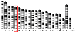
The HTT gene is located on the short arm (p) of chromosome 4 at position 16.3, from base pair 3,074,510 to base pair 3,243,960.[9]
Protein
[edit]Function
[edit]The function of huntingtin (Htt) is not well understood but it is involved in axonal transport.[10] Huntingtin is essential for development, and its absence is lethal in mice.[8] The protein has no sequence homology with other proteins and is highly expressed in neurons and testes in humans and rodents.[11] Huntingtin upregulates the expression of brain-derived neurotrophic factor (BDNF) at the transcription level, but the mechanism by which huntingtin regulates gene expression has not been determined.[12] From immunohistochemistry, electron microscopy, and subcellular fractionation studies of the molecule, it has been found that huntingtin is primarily associated with vesicles and microtubules.[13][14] These appear to indicate a functional role in cytoskeletal anchoring or transport of mitochondria. The Htt protein is involved in vesicle trafficking as it interacts with HIP1, a clathrin-binding protein, to mediate endocytosis, the trafficking of materials into a cell.[15][16] Huntingtin has also been shown to have a role in the establishment in epithelial polarity through its interaction with RAB11A.[17]
Interactions
[edit]Huntingtin has been found to interact directly with at least 19 other proteins, of which six are used for transcription, four for transport, three for cell signalling, and six others of unknown function (HIP5, HIP11, HIP13, HIP15, HIP16, and CGI-125).[18] Over 100 interacting proteins have been found, such as huntingtin-associated protein 1 (HAP1) and huntingtin interacting protein 1 (HIP1), these were typically found using two-hybrid screening and confirmed using immunoprecipitation.[19][20]
| Interacting Protein | PolyQ length dependence | Function |
|---|---|---|
| α-adaptin C/HYPJ | Yes | Endocytosis |
| Akt/PKB | No | Kinase |
| CBP | Yes | Transcriptional co-activator with acetyltransferase activity |
| CA150 | No | Transcriptional activator |
| CIP4 | Yes | cdc42-dependent signal transduction |
| CtBP | Yes | Transcription factor |
| FIP2 | Not known | Cell morphogenesis |
| Grb2[21] | Not known | Growth factor receptor binding protein |
| HAP1 | Yes | Membrane trafficking |
| HAP40 (F8A1, F8A2, F8A3) | Not known | Unknown |
| HIP1 | Yes | Endocytosis, proapoptotic |
| HIP14/HYP-H | Yes | Trafficking, endocytosis |
| N-CoR | Yes | Nuclear receptor co-repressor |
| NF-κB | Not known | Transcription factor |
| p53[22] | No | Transcription factor |
| PACSIN1[23] | Yes | Endocytosis, actin cytoskeleton |
| DLG4 (PSD-95) | Yes | Postsynaptic Density 95 |
| RASA1 (RasGAP)[21] | Not known | Ras GTPase activating protein |
| SH3GL3[24] | Yes | Endocytosis |
| SIN3A | Yes | Transcriptional repressor |
| Sp1[25] | Yes | Transcription factor |
Huntingtin has also been shown to interact with:
Mitochondrial dysfunction
[edit]Huntingtin is a scaffolding protein in the ATM oxidative DNA damage response complex. Mutant huntingtin (mHtt) plays a key role in mitochondrial dysfunction involving the inhibition of mitochondrial electron transport, higher levels of reactive oxygen species and increased oxidative stress.[32][33] The promotion of oxidative damage to DNA may contribute to Huntington's disease pathology.[34]
Clinical significance
[edit]| Repeat count | Classification | Disease status |
|---|---|---|
| <26 | Normal | Unaffected |
| 27–35 | Intermediate | Unaffected |
| 36–40 | Reduced penetrance | +/- Affected |
| >40 | Full penetrance | Affected |
Huntington's disease (HD) is caused by a mutated form of the huntingtin gene, where excessive (more than 36) CAG repeats result in formation of an unstable protein.[35] These expanded repeats lead to production of a huntingtin protein that contains an abnormally long polyglutamine tract at the N-terminus. This makes it part of a class of neurodegenerative disorders known as trinucleotide repeat disorders or polyglutamine disorders. The key sequence which is found in Huntington's disease is a trinucleotide repeat expansion of glutamine residues beginning at the 18th amino acid. In unaffected individuals, this contains between 9 and 35 glutamine residues with no adverse effects.[5] However, 36 or more residues produce an erroneous mutant form of Htt, (mHtt). Reduced penetrance is found in counts 36–39.[36]
Enzymes in the cell often cut this elongated protein into fragments. The protein fragments form abnormal clumps, known as neuronal intranuclear inclusions (NIIs), inside nerve cells, and may attract other, normal proteins into the clumps. The characteristic presence of these clumps in patients was thought to contribute to the development of Huntington disease.[37] However, later research raised questions about the role of the inclusions (clumps) by showing the presence of visible NIIs extended the life of neurons and acted to reduce intracellular mutant huntingtin in neighboring neurons.[38] One confounding factor is that different types of aggregates are now recognised to be formed by the mutant protein, including protein deposits that are too small to be recognised as visible deposits in the above-mentioned studies.[39] The likelihood of neuronal death remains difficult to predict. Likely multiple factors are important, including: (1) the length of CAG repeats in the huntingtin gene and (2) the neuron's exposure to diffuse intracellular mutant huntingtin protein. NIIs (protein clumping) can be helpful as a coping mechanism—and not simply a pathogenic mechanism—to stem neuronal death by decreasing the amount of diffuse huntingtin.[40] This process is particularly likely to occur in the striatum (a part of the brain that coordinates movement) primarily, and the frontal cortex (a part of the brain that controls thinking and emotions).
People with 36 to 40 CAG repeats may or may not develop the signs and symptoms of Huntington disease, while people with more than 40 repeats will develop the disorder during a normal lifetime. When there are more than 60 CAG repeats, the person develops a severe form of HD known as juvenile HD. Therefore, the number of CAG (the sequence coding for the amino acid glutamine) repeats influences the age of onset of the disease. No case of HD has been diagnosed with a count less than 36.[36]
As the altered gene is passed from one generation to the next, the size of the CAG repeat expansion can change; it often increases in size, especially when it is inherited from the father. People with 28 to 35 CAG repeats have not been reported to develop the disorder, but their children are at risk of having the disease if the repeat expansion increases.
References
[edit]- ^ Jump up to: a b c GRCh38: Ensembl release 89: ENSG00000197386 – Ensembl, May 2017
- ^ Jump up to: a b c GRCm38: Ensembl release 89: ENSMUSG00000029104 – Ensembl, May 2017
- ^ "Human PubMed Reference:". National Center for Biotechnology Information, U.S. National Library of Medicine.
- ^ "Mouse PubMed Reference:". National Center for Biotechnology Information, U.S. National Library of Medicine.
- ^ Jump up to: a b The Huntington's Disease Collaborative Research Group (Mar 1993). "A novel gene containing a trinucleotide repeat that is expanded and unstable on Huntington's disease chromosomes. The Huntington's Disease Collaborative Research Group" (PDF). Cell. 72 (6): 971–83. doi:10.1016/0092-8674(93)90585-E. hdl:2027.42/30901. PMID 8458085. S2CID 802885. Archived from the original on 2020-03-13. Retrieved 2019-08-29.
- ^ Choi YB, Kadakkuzha BM, Liu XA, Akhmedov K, Kandel ER, Puthanveettil SV (July 23, 2014). "Huntingtin is critical both pre- and postsynaptically for long-term learning-related synaptic plasticity in Aplysia". PLOS ONE. 9 (7): e103004. Bibcode:2014PLoSO...9j3004C. doi:10.1371/journal.pone.0103004. PMC 4108396. PMID 25054562.
- ^ Nance MA, Mathias-Hagen V, Breningstall G, Wick MJ, McGlennen RC (Jan 1999). "Analysis of a very large trinucleotide repeat in a patient with juvenile Huntington's disease". Neurology. 52 (2): 392–4. doi:10.1212/wnl.52.2.392. PMID 9932964. S2CID 33091017. Archived from the original on 2009-05-05. Retrieved 2009-05-02.
- ^ Jump up to: a b Nasir J, Floresco SB, O'Kusky JR, Diewert VM, Richman JM, Zeisler J, Borowski A, Marth JD, Phillips AG, Hayden MR (Jun 1995). "Targeted disruption of the Huntington's disease gene results in embryonic lethality and behavioral and morphological changes in heterozygotes". Cell. 81 (5): 811–23. doi:10.1016/0092-8674(95)90542-1. PMID 7774020. S2CID 16835259.
- ^ "HTT gene". Archived from the original on 2016-02-02. Retrieved 2016-02-18.
- ^ Vitet H, Brandt V, Saudou F (August 2020). "Traffic signaling: new functions of huntingtin and axonal transport in neurological disease". Current Opinion in Neurobiology. 63: 122–130. doi:10.1016/j.conb.2020.04.001. PMID 32408142. S2CID 218596089.
- ^ Cattaneo E, Zuccato C, Tartari M (December 2005). "Normal huntingtin function: an alternative approach to Huntington's disease". Nature Reviews. Neuroscience. 6 (12): 919–30. doi:10.1038/nrn1806. PMID 16288298. S2CID 10119487.
- ^ Zuccato C, Ciammola A, Rigamonti D, Leavitt BR, Goffredo D, Conti L, et al. (July 2001). "Loss of huntingtin-mediated BDNF gene transcription in Huntington's disease". Science. 293 (5529): 493–8. doi:10.1126/science.1059581. PMID 11408619. S2CID 20703272.
- ^ Hoffner G, Kahlem P, Djian P (March 2002). "Perinuclear localization of huntingtin as a consequence of its binding to microtubules through an interaction with beta-tubulin: relevance to Huntington's disease". Journal of Cell Science. 115 (Pt 5): 941–8. doi:10.1242/jcs.115.5.941. PMID 11870213.
- ^ DiFiglia M, Sapp E, Chase K, Schwarz C, Meloni A, Young C, et al. (May 1995). "Huntingtin is a cytoplasmic protein associated with vesicles in human and rat brain neurons". Neuron. 14 (5): 1075–81. doi:10.1016/0896-6273(95)90346-1. PMID 7748555. S2CID 18071283.
- ^ Velier J, Kim M, Schwarz C, Kim TW, Sapp E, Chase K, et al. (July 1998). "Wild-type and mutant huntingtins function in vesicle trafficking in the secretory and endocytic pathways". Experimental Neurology. 152 (1): 34–40. doi:10.1006/exnr.1998.6832. PMID 9682010. S2CID 36726422.
- ^ Waelter S, Scherzinger E, Hasenbank R, Nordhoff E, Lurz R, Goehler H, et al. (August 2001). "The huntingtin interacting protein HIP1 is a clathrin and alpha-adaptin-binding protein involved in receptor-mediated endocytosis". Human Molecular Genetics. 10 (17): 1807–17. doi:10.1093/hmg/10.17.1807. PMID 11532990.
- ^ Elias S, McGuire JR, Yu H, Humbert S (May 2015). "Huntingtin Is Required for Epithelial Polarity through RAB11A-Mediated Apical Trafficking of PAR3-aPKC". PLOS Biology. 13 (5): e1002142. doi:10.1371/journal.pbio.1002142. PMC 4420272. PMID 25942483.
- ^ Harjes P, Wanker EE (Aug 2003). "The hunt for huntingtin function: interaction partners tell many different stories". Trends in Biochemical Sciences. 28 (8): 425–33. doi:10.1016/S0968-0004(03)00168-3. PMID 12932731.
- ^ Goehler H, Lalowski M, Stelzl U, Waelter S, Stroedicke M, Worm U, Droege A, Lindenberg KS, Knoblich M, Haenig C, Herbst M, Suopanki J, Scherzinger E, Abraham C, Bauer B, Hasenbank R, Fritzsche A, Ludewig AH, Büssow K, Buessow K, Coleman SH, Gutekunst CA, Landwehrmeyer BG, Lehrach H, Wanker EE (Sep 2004). "A protein interaction network links GIT1, an enhancer of huntingtin aggregation, to Huntington's disease". Molecular Cell. 15 (6): 853–65. doi:10.1016/j.molcel.2004.09.016. PMID 15383276.
- ^ Wanker EE, Rovira C, Scherzinger E, Hasenbank R, Wälter S, Tait D, Colicelli J, Lehrach H (Mar 1997). "HIP-I: a huntingtin interacting protein isolated by the yeast two-hybrid system". Human Molecular Genetics. 6 (3): 487–95. doi:10.1093/hmg/6.3.487. PMID 9147654.
- ^ Jump up to: a b Liu YF, Deth RC, Devys D (Mar 1997). "SH3 domain-dependent association of huntingtin with epidermal growth factor receptor signaling complexes". The Journal of Biological Chemistry. 272 (13): 8121–4. doi:10.1074/jbc.272.13.8121. PMID 9079622.
- ^ Steffan JS, Kazantsev A, Spasic-Boskovic O, Greenwald M, Zhu YZ, Gohler H, Wanker EE, Bates GP, Housman DE, Thompson LM (Jun 2000). «Белок болезни Хантингтона взаимодействует с p53 и CREB-связывающим белком и подавляет транскрипцию» . Труды Национальной академии наук Соединенных Штатов Америки . 97 (12): 6763–8. Bibcode : 2000pnas ... 97.6763s . doi : 10.1073/pnas.100110097 . PMC 18731 . PMID 10823891 .
- ^ Modregger J, Diprospero NA, Charles V, Tagle DA, Plomann M (октябрь 2002 г.). «Pacsin 1 взаимодействует с Huntingtin и отсутствует в синаптических варикозах в мозгах болезней -болезней Хантингтона» . Молекулярная генетика человека . 11 (21): 2547–58. doi : 10.1093/hmg/11.21.2547 . PMID 12354780 .
- ^ Sittler A, Wälter S, Wedemeyer N, Hasenbank R, Scherzinger E, Eickhoff H, Bates GP, Lehrach H, Wanker EE (октябрь 1998). «SH3GL3 ассоциируется с белком Huntingtin Exon 1 и способствует образованию полиглинсодержащих белковых агрегатов» . Молекулярная клетка . 2 (4): 427–36. doi : 10.1016/s1097-2765 (00) 80142-2 . PMID 9809064 .
- ^ Ли Ш., Ченг А.Л., Чжоу Х., Лам С., Рао М., Ли Х, Ли XJ (март 2002 г.). «Взаимодействие белка болезни Хантингтона с транскрипционным активатором SP1» . Молекулярная и клеточная биология . 22 (5): 1277–87. doi : 10.1128/mcb.22.5.1277-1287.2002 . PMC 134707 . PMID 11839795 .
- ^ Калхман М.А., Грэм Р.К., Ся Г., Коид Х.Б., Ходжсон Дж.Г., Грэм К.С., Голдберг Ю.П., Гитц Р.Д., Пикарт С.М., Хейден М.Р. (август 1996 г.). «Хантингтин убиквитинирован и взаимодействует с конкретным убиквитином, конъюгирующим фермент» . Журнал биологической химии . 271 (32): 19385–94. doi : 10.1074/jbc.271.32.19385 . PMID 8702625 .
- ^ Лю Иф, Дороу Д., Маршалл Дж. (Jun 2000). «Активация MLK2-опосредованных сигнальных каскадов с помощью Huntingtin, проведенного с полиглутамином» . Журнал биологической химии . 275 (25): 19035–40. doi : 10.1074/jbc.c000180200 . PMID 10801775 .
- ^ Hattula K, Peränen J (2000). «FIP-2, белок с спиральной катушкой, связывает хантингтин с Rab8 и модулирует клеточный морфогенез» . Текущая биология . 10 (24): 1603–6. doi : 10.1016/s0960-9822 (00) 00864-2 . PMID 11137014 . S2CID 12836037 .
- ^ Jump up to: а беременный в Faber PW, Barnes GT, Srinidhi J, Chen J, Gusella JF, Macdonald ME (сентябрь 1998). «Хантингтин взаимодействует с семейством белков домена WW» . Молекулярная генетика человека . 7 (9): 1463–74. doi : 10.1093/hmg/7.9.1463 . PMID 9700202 .
- ^ Холберт С., Дедеоглу А., Гумберт С., Сауду Ф., Ферранте Р.Дж., Нери С (март 2003 г.). «CDC42-взаимодействующий белок 4 связывается с хантингтином: невропатологические и биологические данные о роли в болезни Хантингтона» . Труды Национальной академии наук Соединенных Штатов Америки . 100 (5): 2712–7. Bibcode : 2003pnas..100.2712H . doi : 10.1073/pnas.0437967100 . PMC 151406 . PMID 12604778 .
- ^ Сингараджа Р.Р., Хадано С., Метцлер М., Гиван С., Веллингтон К.Л., Варби С., Янай А., Гутекунст К.А., Ливитт Б.Р., Йи Х, Фихтер К., Ган Л., Маккатчон К., Чопра В., Мишель Дж., Герш С.М., Икеда Дж. , Хейден М.Р. (ноябрь 2002 г.). «HIP14, новый домен анкиринового домена, связывает хантингтин с внутриклеточным переносом и эндоцитозом» . Молекулярная генетика человека . 11 (23): 2815–28. doi : 10.1093/hmg/11.23.2815 . PMID 12393793 .
- ^ Liu Z, Zhou T, Ziegler AC, Dimitrion P, Zuo L (2017). «Окислительный стресс при нейродегенеративных заболеваниях: от молекулярных механизмов до клинических применений» . Оксидные медные ячейки Longev . 2017 : 2525967. DOI : 10.1155/2017/2525967 . PMC 5529664 . PMID 28785371 .
- ^ Майури, Тамара; Мокл, Эндрю Дж.; Хунг, Клаудия Л.; Ся, Цзяньрун; Ван Рон-мам, Вилле М.К.; Острый, Рэй (25 декабря 2016 г.). «Хантингтин - это белок каркасов в комплексе ответа на окислительный повреждение окислительного повреждения АТР» » . Молекулярная генетика человека . 26 (2): 395–406. doi : 10.1093/hmg/ddw395 . PMID 28017939 .
- ^ Ayala-Peña S (сентябрь 2013 г.). «Роль окислительного повреждения ДНК в митохондриальной дисфункции и патогенеза болезни Хантингтона» . Свободный радик. Биол. Медик 62 : 102–10. doi : 10.1016/j.freeradbiomed.2013.04.017 . PMC 3722255 . PMID 23602907 .
- ^ Jump up to: а беременный Уокер Ф. Ф. (январь 2007 г.). «Болезнь Хантингтона». Лансет . 369 (9557): 218–28. doi : 10.1016/s0140-6736 (07) 60111-1 . PMID 17240289 . S2CID 46151626 .
- ^ Jump up to: а беременный Chong SS, Almqvist E, Telenius H, Latray L, Nichol K, Bourdelat-Parks B, Goldberg YP, Haddad BR, Richards F, Sillence D, Greenberg CR, Ives E, Van Dengh G, Hughes MR, Hayden Mr (Feb 1997). «Вклад последовательности ДНК и размера CAG в частоты мутаций промежуточных аллелей при заболевании Хантингтона: данные анализа отдельных сперматозоидов» . Молекулярная генетика человека . 6 (2): 301–9. doi : 10.1093/hmg/6.2.301 . PMID 9063751 .
- ^ Davies SW, Turmaine M, Cozens BA, Difiglia M, Sharp AH, Ross CA, Scherzinger E, Wanker EE, Mangiarini L, Bates GP (август 1997 г.). «Образование нейрональных внутриядерных включений лежит в основе неврологической дисфункции у мышей, трансгенных для мутации HD» . Клетка . 90 (3): 537–48. doi : 10.1016/s0092-8674 (00) 80513-9 . PMID 9267033 . S2CID 549691 .
- ^ Arrasate M, Mitra S, Schweitzer ES, Segal MR, Finkbeiner S (октябрь 2004 г.). «Образование тела включения снижает уровни мутанта -хантингтина и риск гибели нейронов» . Природа . 431 (7010): 805–10. Bibcode : 2004natur.431..805a . doi : 10.1038/nature02998 . PMID 15483602 .
- ^ Sahl SJ, Lau L, Vonk WI, Weiss Le, Frydman J, Moerner WE (2016). «Задержка появления фибриллов мутантов размером размером с субдифракции после образования тела включения» . Q Rev Biophys . 49 : E2. doi : 10.1017/s0033583515000219 . PMC 4785097 . PMID 26350150 .
- ^ Orr ht (октябрь 2004 г.). «Нейродегенеративное заболевание: Агентство по защите нейронов». Природа . 431 (7010): 747–8. Bibcode : 2004natur.431..747o . doi : 10.1038/431747a . PMID 15483586 . S2CID 285829 .
Дальнейшее чтение
[ редактировать ]- Kosinski CM, Schlangen C, Gellerich FN, Gizatullina Z, Deschauer M, Schiefer J, et al. (Август 2007 г.). «Миопатия как первый симптом болезни Хантингтона в марафонском бегуне». Движение расстройства . 22 (11): 1637–40. doi : 10.1002/mds.21550 . PMID 17534945 . S2CID 30904037 .
- Бейтс G (май 2003 г.). «Агрегация и токсичность Хантингтина при болезни Хантингтона». Лансет . 361 (9369): 1642–4. doi : 10.1016/s0140-6736 (03) 13304-1 . PMID 12747895 . S2CID 7587406 .
- Cattaneo E (февраль 2003 г.). «Дисфункция хантингтина дикого типа при болезни Хантингтона». Новости в физиологических науках . 18 : 34–7. doi : 10.1152/nips.01410.2002 . PMID 12531930 .
- Gárdián G, Vécsei L (октябрь 2004 г.). «Болезнь Хантингтона: патомеханизм и терапевтические перспективы». Журнал нейронной передачи . 111 (10–11): 1485–94. doi : 10.1007/s00702-004-0201-4 . PMID 15480847 . S2CID 2961376 .
- Landles C, Bates GP (октябрь 2004 г.). «Хантингтин и молекулярный патогенез болезни Хантингтона. Четвертый в серии обзоров молекулярной медицины» . Embo сообщает . 5 (10): 958–63. doi : 10.1038/sj.embor.7400250 . PMC 1299150 . PMID 15459747 .
- Джонс Ал (июнь 1999). «Локализация и взаимодействие Хантингтина» . Философские транзакции Королевского общества Лондона. Серия B, биологические науки . 354 (1386): 1021–7. doi : 10.1098/rstb.1999.0454 . PMC 1692601 . PMID 10434301 .
- Ли Ш., Ли XJ (октябрь 2004 г.). «Хантингтин и его роль в дегенерации нейронов». Нейробиолог . 10 (5): 467–75. doi : 10.1177/10738584042667777 . PMID 15359012 . S2CID 19491573 .
- MacDonald ME, Novelletto A, Lin C, Tagle D, Barnes G, Bates G, Taylor S, Allitto B, Altherr M, Myers R (май 1992). «В регионе кандидата в болезнь Хантингтона есть много разных гаплотипов». Природа генетика . 1 (2): 99–103. doi : 10.1038/ng0592-99 . PMID 1302016 . S2CID 25472459 .
- Макдональд меня (ноябрь 2003 г.). «Хантингтин: жив и хорошо и работает в среднем управлении». Science's Stke . 2003 (207): PE48. doi : 10.1126/stke.2003.207.pe48 . PMID 14600292 . S2CID 35318234 .
- Майерс Р.Х. (апрель 2004 г.). «Генетика болезни Хантингтона» . Нейрорус . 1 (2): 255–62. doi : 10.1602/neuroRx.1.2.255 . PMC 534940 . PMID 15717026 .
- Рангоне Х, Гумберт С., Сауду Ф (июль 2004). «Болезнь Хантингтона: как Хантингтин, антиапоптотический белок, становится токсичным?». Патология-биология . 52 (6): 338–42. doi : 10.1016/j.patbio.2003.06.004 . PMID 15261377 .
- Молодой AB (февраль 2003 г.). «Хантингтин в здоровье и болезнях» . Журнал клинических исследований . 111 (3): 299–302. doi : 10.1172/jci17742 . PMC 151871 . PMID 12569151 .
Внешние ссылки
[ редактировать ]- Хантингтин+белок,+человек в Национальной библиотеке медицины США. Медицинские заголовки (сетка)
- Агрегация белка и белка Хантингтина в надежде, архивные 2021-02-12 на The Wayback Machine : Huntington's Outreach Project по образованию в Стэнфорде
- Ассоциация болезни HDA Huntington UK
- Онлайн -наследство Менделян в Человеке (Омим): 143100
- Entergene 3064
- Genecard
- Вместе