Стоматологическая травма
| Стоматологическая травма | |
|---|---|
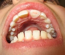 | |
| Сломанный верхний передний зуб. Слои ткани, которые составляют зуб, четко видны, с розовой мякотью, выделяющейся на более дорогой дентине и эмале зуба . | |
| Специальность | Пероральная и челюстная хирургия |
Травма зубов относится к травме (травмам) зубам и /или периодонту ( десен , пародонта, связки , альвеолярная кость ) и близлежащие мягкие ткани , такие как губы, язык и т. Д. [ 1 ]
Типы
[ редактировать ]

Стоматологические травмы
[ редактировать ]Стоматологические травмы включают: [ 2 ] [ 3 ]
- Нарушение эмали
- Эмаль перелом
- Эмаль-Динтин перелома
- Перелом эмали-дентин, включающий воздействие мякоти
- Корневая перелома зуба
Периодонтальные травмы
[ редактировать ]- Сотрясение мозга (синяки)
- Подвижность зуба (зуб сбил свободу)
- Вывитие зуба (перемещение)
- Экстремивный
- Навязчивый
- Боковой
- Авульсия зуба [ 4 ] (Зуб нокаутировал)
Травмы поддержки кости
[ редактировать ]
Эта травма включает в себя альвеолярную кость и может выходить за пределы альвеола. [ 5 ] [ 6 ] Существует пять различных типов альвеолярных переломов:
- Общается перелом стены гнезда
- Перелом стены гнезда
- Зубчатый перелом (сегментарный)
- Перелом верхней челюсти: перелом в Форт , зигоматический перелом , орбитальный прорыв
- Перелом нижней челюсти
Повреждения травмы, связанные с альвеолом, могут быть сложными, поскольку это не происходит в изоляции, очень часто представляет собой другие виды травм зубной ткани.
Признаки зубчатого перелома:
- Изменение на окклюзию
- Несколько зубов движутся вместе как сегмент и обычно смещены
- Синяки прикрепленного десны
- Десны по линии перелома часто разорваны
Исследование : требуется более чем одно рентгенографическое представление для определения линии перелома.
Обработка : переназначенная перемещение зубов под локальным анестетиком и стабилизируйте подвижный сегмент шинкой в течение 4 недель, швов любые раны мягких тканей.
Разрыв мягких тканей
[ редактировать ]
Повреждения мягких тканей обычно представлены в связи с стоматологической травмой. Обычно затрагиваемые участки - губы, слизистая оболочка щека, десны, френа и язык. Наиболее распространенными травмами являются губы и гингива. Для губ важно исключить присутствие посторонних предметов в ранах и рваных разрывах путем тщательного изучения. Рентгенограмма может быть взята для выявления любых потенциальных посторонних объектов. [ 5 ] [ 6 ]
Руки Gingivae, которые являются небольшими, обычно заживают спонтанно и не требуют какого -либо вмешательства. Однако это может быть одной из клинических представлений альвеолярного перелома. Гингива, кровоточащая, особенно вокруг краев, может указывать на повреждение пародонтальной связки зуба.
Лицевой нерв и позвоночный проток следует исследовать на предмет любого потенциального повреждения, когда участвует слизистая оболочка щека.
Глубокие раны ткани должны быть восстановлены в слоях с помощью резорбируемых швов.
Первичные зубы
[ редактировать ]Травма первичных зубов чаще всего в возрасте от двух до трех лет во время развития моторной координации. [ 7 ] Когда первичные зубы получают травмы, полученное лечение приоритет в безопасности взрослого зуба, [ 7 ] и следует избегать любого риска повреждения постоянных преемников. [ 8 ] Это связано с тем, что корневая вершина поврежденного первичного зуба находится вблизи зародыша зуба взрослого зуба. [ 8 ]
Следовательно, первичный первичный зуб будет удален, если будет обнаружено, что он вторгся на развивающуюся зародышевой зародышевой зародыши для взрослых. [ 8 ] Если это произойдет, родителям следует посоветовать о возможных осложнениях, таких как гипоплазия эмали, гипокальцификация , расширение коронки/корня или нарушения в последовательности извержения зубов. [ 9 ]
Potential sequelae can involve pulpal necrosis, pulp obliteration and root resorption.[10] Necrosis is the most common complication and an assessment is generally made based on the colour supplemented with radiograph monitoring. A change in colour may mean that the tooth is still vital but if this persists it is likely to be non-vital.
Permanent teeth
[edit]Dental injuries
[edit]| Dental injury[2] | Clinical findings | Radiographic findings | Treatment | Follow-up |
|---|---|---|---|---|
| 1) Enamel infraction | A crack in enamel with no loss of tooth structure
Tooth is not tender |
No abnormalities
|
Generally no treatment needed
Discolouration of prominent cracks can be prevented by etching and sealing with resin |
No follow-up required |
| 2) Enamel fracture | Fracture involving enamel only
Tooth not tender with normal mobility and pulpal response |
Loss of enamel
May need lip or cheek x-ray to locate tooth fragments or other materials
|
If available, tooth fragment can be bonded back onto the tooth
If not available, tooth can be restored with composite resin |
6–8 weeks: clinical and radiographic examination
1 year: clinical and radiographic examination |
| 3) Enamel-dentine fracture | Fracture involving both enamel and dentine without pulp exposure
Tooth not tender with normal mobility and pulpal response |
Loss of enamel and dentine
May need lip or cheek x-ray to locate tooth fragments or other materials |
If available, tooth fragment can be bonded back onto the tooth
If not available, tooth can be restored with composite resin If dentine is within 0.5mm of the pulp, calcium hydroxide placed and covered with glass ionomer |
6–8 weeks: clinical and radiographic examination
1 year: clinical and radiographic examination |
| 4) Enamel-dentine-pulp fracture | Fracture involving enamel and dentine with pulp exposure
Tooth not tender with normal mobility Exposed pulp will be sensitive to stimuli |
Loss of enamel and dentine
May need lip or cheek x-ray to locate tooth fragments or other materials
|
In developing teeth, preserve pulp vitality by pulp capping or partial pulpotomy using calcium hydroxide
In mature teeth, root canal treatment is usually performed |
6–8 weeks: clinical and radiographic examination
1 year: clinical and radiographic examination |
| 5) Crown-root fracture without pulp involvement | Fracture involving enamel, dentine and cementum without pulp exposure
Fracture extends below the gum margin Tender tooth with mobile crown fragment |
Fracture line extending down the root may not be visible | Emergency:
aim is to stabilise the loose fragment by splinting it to adjacent teeth Non-emergency: removal of loose fragment (following gingivectomy, surgery or via orthodontics), root canal treatment and restoration with post-retained crown In extreme cases (such as a vertical fracture), tooth may need to be extracted |
6–8 weeks: clinical and radiographic examination
1 year: clinical and radiographic examination |
| 6) Crown-root fracture with pulp Involvement | Fracture involving enamel, dentine and cementum with pulp exposure
Tender tooth with mobile crown fragment |
Fracture line extending down the root may not be visible | Emergency:
aim is to stabilise the loose fragment by splinting it to adjacent teeth In developing teeth, preserve pulp vitality by pulp capping or partial pulpotomy using calcium hydroxide In mature teeth, root canal treatment is usually performed Non-emergency: removal of loose fragment (following gingivectomy, surgery or via orthodontics), root canal treatment and restoration with post-retained crown In extreme cases (such as a vertical fracture), tooth may need to be extracted |
6–8 weeks: clinical and radiographic examination
1 year: clinical and radiographic examination |
| 7) Root fracture | Mobile or displaced crown segment
Tender tooth that may be bleeding from the gum Tooth may be discoloured (red or grey) |
Fracture line involving the root will be seen as well as the direction | If displaced, reposition tooth and check the position with an x-ray
Flexible splint used to stabilise tooth for at least 4 weeks and then reassess tooth stability Monitor healing for at least 1 year to assess the status of the pulp Root canal treatment will be needed if pulp necrosis develops (this occurs in ~20% of root fractures) |
4 weeks: splint removal, clinical and radiographic examination
6–8 weeks: clinical and radiographic examination 4 months: splint removal in cervical third fractures, clinical and radiographic examination 6 months: clinical and radiographic examination 1 year: clinical and radiographic examination 5 years: clinical and radiographic examination |
Periodontal injuries
[edit]| Periodontal injury[2] | Clinical findings | Radiographic findings | Treatment | Follow-up |
|---|---|---|---|---|
| 1) Concussion | Tender tooth with no displacement and normal mobility | No abnormalities | No treatment required | 4 weeks: clinical and radiographic examination
6–8 weeks: clinical and radiographic examination 1 year: clinical and radiographic examination |
| 2) Subluxation | Tender tooth with no displacement but increased mobility
May be bleeding from the gum |
No abnormalities | Usually no treatment required
Can use a flexible splint to stabilise the tooth for up to 2 weeks |
2 weeks: splint removal, clinical and radiographic examination
4 weeks: clinical and radiographic examination 6–8 weeks: clinical and radiographic examination 6 months: clinical and radiographic examination 1 year: clinical and radiographic examination |
| 3) Extrusion | Tooth looks longer and is very mobile | Periodontal ligament space is increased apically | Tooth is repositioned gently in the socket
Tooth stabilised with a flexible splint for 2 weeks Signs and symptoms of pulp necrosis indicates the need for root canal treatment to prevent root resorption |
2 weeks: splint removal, clinical and radiographic examination
4 weeks: clinical and radiographic examination 6–8 weeks: clinical and radiographic examination 6 months: clinical and radiographic examination 1 year: clinical and radiographic examination yearly 5 years: clinical and radiographic examination |
| 4) Lateral luxation | Tooth is displaced, most commonly towards the roof of the mouth/tongue or the lip
Tooth will be immobile Tapping on the tooth will give a high-pitched, metallic (ankylotic) sound Alveolar process fracture |
Periodontal ligament space is widened | Reposition the tooth using fingers or forceps to remove its "bony lock" and gently reposition it in the socket
Tooth stabilised with a flexible splint for 4 weeks Signs and symptoms of pulp necrosis indicates the need for root canal treatment to prevent root resorption |
2 weeks: clinical and radiographic examination
4 weeks: splint removal, clinical and radiographic examination 6–8 weeks: clinical and radiographic examination 6 months: clinical and radiographic examination 1 year: clinical and radiographic examination Yearly for 5 years: clinical and radiographic examination |
| 5) Intrusion | Tooth is displaced into the alveolar bone
Tapping on the tooth will give a high-pitched, metallic (ankylotic) sound |
Absence of periodontal ligament space from part or entirety of the root
Cemento-enamel junction appears more apically for the traumatised tooth |
If incomplete root formation:
allow time for tooth to naturally erupt but if no movement after a few weeks then start orthodontic repositioning intruded >7mm: surgical or orthodontic repositioning stabilise with flexible splint for 4 weeks after repositioning If complete root formation: intruded <3mm: allow time for natural eruption but if no movement after 2–4 weeks then reposition surgically or orthodontically intruded 3-7mm: surgical or orthodontic repositioning intruded >7mm: surgical repositioning likelihood of pulpal necrosis in these teeth so root canal therapy with temporary calcium hydroxide filling in first instance and start root canal treatment 2–3 weeks after repositioning stabilise with flexible splint for 4 weeks after repositioning |
2 weeks: clinical and radiographic examination
4 weeks: splint removal, clinical and radiographic examination 6–8 weeks: clinical and radiographic examination 6 months: clinical and radiographic examination 1 year: clinical and radiographic examination Yearly for 5 years: clinical and radiographic examination |
| 6) Avulsion | Tooth completely removed from socket | Radiograph required to ensure that missing tooth is not intruded | For information on first aid procedures for avulsed teeth, see "Management" section of this page
Treatment will depend on whether the tooth has an open or closed apex and how long the tooth has been out of the mouth prior to dental clinic arrival (see Dental Trauma Guide for full treatment details) |
4 weeks: splint removal, clinical and radiographic examination
3 months: clinical and radiographic examination 6 months: clinical and radiographic examination 1 year: clinical and radiographic examination yearly |
Risk factors
[edit]- Age, especially young children[8][9][11]
- Primary dentition stage (2–3 years old, when children's motor function is developing and start learning how to walk/ run)
- Mixed dentition stage (8–10 years old)
- Permanent dentition stage (13–15 years old)
- Male > Female [11][12]
- Season (Many trauma incidents occur more in summer compared to winter)[13]
- Sports, especially contact sports such as football, hockey, rugby, basketball and skating [14]
- Piercing in tongue and lips[15][16][17]
- Military training[18][19]
- Acute changes in the barometric pressure, i.e. dental barotrauma,[20] which can affect scuba divers[21] and aviators[22]
- Class II malocclusion with increased overjet and Class II skeletal relationship [23][24] and incompetent lips[25] are the significant risk factors
Prevention
[edit]Prevention in general is relatively difficult as it is nearly impossible to stop accidents from happening, especially in children who are quite active. Regular use of a gum shield during sports and other high-risk activities (such as military training) is the most effective prevention for dental trauma.[26][27] They are mainly being fitted on the upper teeth as it has higher risk of dental trauma compared to the lower teeth. Gum shields ideally have to be comfortable for users, retentive, odourless, tasteless and the materials should not be causing any harm to the body.[28] However, studies in various high-risk populations for dental injuries have repeatedly reported low compliance of individuals for the regular using of mouthguard during activities.[29] Moreover, even with regular use, effectiveness of prevention of dental injuries is not complete, and injuries can still occur even when mouthguards are used as users are not always aware of the best makes or size, which inevitably result in a poor fit.[18]
- Stock ready-moulded
- Not recommended as it does not conform the teeth at all
- Poor retention
- Poor fit
- Higher risk of dislodging during contact sports and airway occlusion which may lead to respiratory distress
- Self-moulded/Boil and bite
- Limited range of sizes, which may result in poor fitting
- Can be easily remoulded if distorted
- Cheap
- Custom-made
- Made with ethylene vinyl acetate
- The most ideal type of gum shield[31]
- Good retention
- Able to build in multiple layers/laminations
- Expensive
One of the most important measures is to impart knowledge and awareness about dental injury to those who are involved in sports environments like boxing and in school children in which they are at high risk of suffering dental trauma through an extensive educational campaign including lectures, leaflets, posters which should be presented in an easy understandable way.[32]
Management
[edit]The management depends on the type of injury involved and whether it is a baby or an adult tooth. If teeth are completely knocked out baby front teeth should not be replaced. The area should be cleaned gently and the child brought to see a dentist. Adult front teeth (which usually erupt at around six years of age) can be replaced immediately if clean. If a tooth is avulsed, make sure it is a permanent tooth (primary teeth should not be replanted, and instead the injury site should be cleaned to allow the adult tooth to begin to erupt).
- Reassure the patient and keep them calm.
- If the tooth can be found, pick it up by the crown (the white part). Avoid touching the root part.
- If the tooth is dirty, wash it briefly (ten seconds) under cold running water but do not scrub the tooth.
- Place the tooth back in the socket where it was lost from, taking care to place it the correct way (matching the other tooth)
- Encourage the patient to bite on a handkerchief to hold the tooth in position.
- If it is not possible to replace the tooth immediately, ideally, the tooth should be placed in Hank's balanced salt solution,[33] if not available, in a glass of milk or a container with the patient's saliva or in the patient's cheek (keeping it between the teeth and the inside of the cheek – note this is not suitable for young children who may swallow the tooth). Transporting the tooth in water is not recommended, as this will damage the delicate cells that make up the tooth's interior.
- Seek emergency dental treatment immediately.
When the injured teeth are painful while functioning due to damage to the periodontal ligaments (e.g., dental subluxation), a temporary splinting of the injured teeth may relieve the pain and enhance eating ability.[34] Splinting should only be used in certain situations. Splinting in lateral and extrusive luxation had a poorer prognosis than in root fractures.[35] An avulsed permanent tooth should be gently rinsed under tap water and immediately re-planted in its original socket within the alveolar bone and later temporarily splinted by a dentist.[4] Failure to re-plant the avulsed tooth within the first 40 minutes after the injury may result in very poor prognosis for the tooth.[4] Management of injured primary teeth differs from management of permanent teeth; an avulsed primary tooth should not be re-planted (to avoid damage to the permanent dental crypt).[8] This is due to the close proximity of the apex of a primary tooth to the permanent tooth underneath. The permanent dentition can suffer from tooth malformation, impacted teeth and eruption disturbances due to trauma to primary teeth. The priority should always be reducing potential damage to the underlying permanent dentition.[36]
For other injuries, it is important to keep the area clean by using a soft toothbrush and antiseptic mouthwash such as chlorhexidine gluconate. Soft foods and avoidance of contact sports is also recommended in the short term. Dental care should be sought as quickly as possible.
Splinting
[edit]A tooth that has experienced trauma may become loose due to the periodontal ligament becoming damaged or fracture to the root of the tooth. Splinting ensures that the tooth is held in the correct position within the socket, ensuring that no further trauma occurs to enable healing.[37] A splint can either be flexible or rigid. Flexible splints do not completely immobilise the traumatised tooth and still allow for functional movement. Contrastingly, rigid splints completely immobilise the traumatised tooth.[38] The International Association of Dental Traumatology (IADT) guidelines recommend the use of flexible, non-rigid splints for a short duration by stating that both periodontal and pulpal healing is encouraged if the traumatised tooth is allowed slight movement and if the splinting time is not too long.[39][40]
Complications
[edit]Not all sequelae of trauma are immediate and many of them can occur months or years after the initial incident thus required prolonged follow-up. Common complications are pulpal necrosis, pulpal obliteration, root resorption and damage to the successors teeth in primary teeth dental trauma. The most common complication was pulp necrosis (34.2%). 50% of the tooth that have trauma related to avulsion experienced ankylotic root resorption after a median TIC (time elapsed between the traumatic event and the diagnosis of complications) of 1.18 years. Teeth that have multiple traumatic events also showed to have higher chance of pulp necrosis (61.9%) compared to teeth that experienced a single traumatic injury (25.3%) in the studies (1)[41]

Pulpal necrosis
[edit]Pulp necrosis usually occurs either as ischaemic necrosis (infarction) caused by disruption to the blood supply at the apical foramen or as an infection-related liquefactive necrosis following dental trauma (2). Signs of pulpal necrosis include[42]
- Persistent grey colour to tooth that does not fade
- Radiographic signs of periapical inflammation
- Clinical signs of infection: tenderness, sinus, suppuration, swelling
Treatment options will be extraction for the primary tooth. For the permanent tooth, endodontic treatment can be considered.

Root resorption
[edit]Root resorption following traumatic dental injuries, whether located along the root surface or within the root canal appears to be a sequel to wound healing events, where a significant amount of the PDL or pulp has been lost due to the effect of acute trauma.[43]
Pulpal obliteration
[edit]4–24% of traumatized teeth will have some degrees of pulpal obliteration that is characterized by the loss of pulpal space radiographically and yellow discolouration of the clinical crown. No treatment is needed if it is asymptomatic. Treatment options will be extraction for symptomatic primary tooth. For symptomatic permanent tooth, root canal treatment is often challenging because the pulp chamber is filled with calcified material and the drop-off sensation of entering a pulp chamber will not occur.[44]
Damage to the successor teeth
[edit]Dental trauma to the primary teeth might cause damage to the permanent teeth. Damage to the permanent teeth especially during development stage might have following consequences:[45]
- Crown dilaceration
- Odontoma-like malformation
- Sequestration of permanent tooth germs
- Root dilaceration
- Arrest of root formation
Epidemiology
[edit]Dental trauma is most common in younger people, accounting for 17% of injuries to the body in those aged 0–6 years compared to an average of 5% across all ages.[46] It is more frequently observed in males compared to females.[47] Traumatic dental injuries are more common in permanent teeth compared to deciduous teeth and usually involve the front teeth of the upper jaw.[48]
"The oral region comprises 1% of the total body area, yet it accounts for 5% of all bodily injuries. In preschool children, oral injuries make up as much as 17% of all bodily injuries. The incidence of traumatic dental injuries is 1–3%, and the prevalence is steady at 20–30%."[49]
Almost 30% of the children in pre-school have mostly experienced trauma to primary teeth. Dental injuries involving the permanent teeth happen to almost 25% of children in school and 30% of adults. The incident varies in different countries as well as within the country itself. Dental traumatic accidents depends on one's activity status and also the surrounding environment factor but these are the main predisposing risk factor compared to a person's age and gender.[50]
Trauma is the most common cause of loss of permanent incisors in childhood. Dental trauma often leads to complications such as pulpal necrosis, and it is nearly impossible to predict the long-term prognosis of the injured tooth; the injury often results in long-term restorative problems.[51][52][53]
See also
[edit]References
[edit]- ^ Textbook and Color Atlas of Traumatic Injuries to the Teeth, Fourth Edition, edited by Andreason J, Andreasen F, and Andersson L, Wiley-Blackwell, Oxford, UK, 2007
- ^ Jump up to: a b c "Permanent teeth – Dental Trauma Guide". Retrieved 2019-01-20.
- ^ "Primary teeth – Dental Trauma Guide". Retrieved 2019-01-20.
- ^ Jump up to: a b c Flores MT, Andersson L, Andreasen JO, Bakland LK, Malmgren B, Barnett F, Bourguignon C, DiAngelis A, Hicks L, Sigurdsson A, Trope M, Tsukiboshi M, von Arx T (June 2007). "Guidelines for the management of traumatic dental injuries. II. Avulsion of permanent teeth". Dental Traumatology. 23 (3): 130–6. doi:10.1111/j.1600-9657.2007.00605.x. PMID 17511833.
- ^ Jump up to: a b Tagar H, Djemal S (September 2017). "Oral surgery II: Part 1. Acute management of dentoalveolar trauma". British Dental Journal. 223 (6): 407–416. doi:10.1038/sj.bdj.2017.805. PMID 28937097. S2CID 205668709.
- ^ Jump up to: a b Durham J, Moore UJ, Hill CM, Renton T (December 2017). "Oral surgery II: Part 6. Oral and maxillofacial trauma". British Dental Journal. 223 (12): 877–883. doi:10.1038/sj.bdj.2017.995. PMID 29269898. S2CID 19070108.
- ^ Jump up to: a b Flores, Marie Therese (2002). "Traumatic injuries in the primary dentition". Dental Traumatology. 18 (6): 287–298. doi:10.1034/j.1600-9657.2002.00153.x. ISSN 1600-4469. PMID 12656861.
- ^ Jump up to: a b c d e Flores MT, Malmgren B, Andersson L, Andreasen JO, Bakland LK, Barnett F, Bourguignon C, DiAngelis A, Hicks L, Sigurdsson A, Trope M, Tsukiboshi M, von Arx T (August 2007). "Guidelines for the management of traumatic dental injuries. III. Primary teeth". Dental Traumatology. 23 (4): 196–202. doi:10.1111/j.1600-9657.2007.00627.x. PMID 17635351.
- ^ Jump up to: a b "Guideline on Management of Acute Dental Trauma" (PDF). Council on Clinical Affairs. 2011.
- ^ Welbury RR, Duggal MS, Hosey MT, eds. (2007). Paediatric Dentistry (Third ed.). Oxford, UK: Oxford University Press. ISBN 978-0071445085.
- ^ Jump up to: a b Jesus MA, Antunes LA, Risso P, Freire MV, Maia LC (January 2010). "Epidemiologic survey of traumatic dental injuries in children seen at the Federal University of Rio de Janeiro, Brazil". Brazilian Oral Research. 24 (1): 89–94. doi:10.1590/S1806-83242010000100015. PMID 20339720.
- ^ Ivancic Jokic N, Bakarcic D, Fugosic V, Majstorovic M, Skrinjaric I (February 2009). "Dental trauma in children and young adults visiting a University Dental Clinic". Dental Traumatology. 25 (1): 84–7. doi:10.1111/j.1600-9657.2008.00711.x. PMID 19208016.
- ^ Perez R, Berkowitz R, McIlveen L, Forrester D (October 1991). "Dental trauma in children: a survey". Endodontics & Dental Traumatology. 7 (5): 212–3. doi:10.1111/j.1600-9657.1991.tb00438.x. PMID 1687388.
- ^ Young EJ, Macias CR, Stephens L (May 2015). "Common Dental Injury Management in Athletes". Sports Health. 7 (3): 250–5. doi:10.1177/1941738113486077. PMC 4482297. PMID 26131303.
- ^ Levin L, Zadik Y, Becker T (December 2005). "Oral and dental complications of intra-oral piercing". Dental Traumatology. 21 (6): 341–3. doi:10.1111/j.1600-9657.2005.00395.x. PMID 16262620.
- ^ Leichter, Jonathan W.; Monteith, Brian D. (February 2006). "Prevalence and risk of traumatic gingival recession following elective lip piercing". Dental Traumatology. 22 (1): 7–13. doi:10.1111/j.1600-9657.2006.00332.x. ISSN 1600-4469. PMID 16422752.
- ^ Hennequin-Hoenderdos, Nl; Slot, De; Van der Weijden, Ga (February 2016). "The incidence of complications associated with lip and/or tongue piercings: a systematic review". International Journal of Dental Hygiene. 14 (1): 62–73. doi:10.1111/idh.12118. ISSN 1601-5029. PMID 25690049.
- ^ Jump up to: a b Zadik Y, Levin L (December 2008). "Orofacial injuries and mouth guard use in elite commando fighters". Military Medicine. 173 (12): 1185–7. doi:10.7205/milmed.173.12.1185. PMID 19149336.
- ^ Zadik Y, Levin L (February 2009). "Oral and facial trauma among paratroopers in the Israel Defense Forces". Dental Traumatology. 25 (1): 100–2. doi:10.1111/j.1600-9657.2008.00719.x. PMID 19208020.
- ^ Zadik Y (Jul–Aug 2009). "Dental barotrauma". The International Journal of Prosthodontics. 22 (4): 354–7. PMID 19639071.
- ^ Zadik Y, Drucker S (September 2011). "Diving dentistry: a review of the dental implications of scuba diving". Australian Dental Journal. 56 (3): 265–71. doi:10.1111/j.1834-7819.2011.01340.x. PMID 21884141.
- ^ Zadik Y (January 2009). "Aviation dentistry: current concepts and practice". British Dental Journal. 206 (1): 11–6. doi:10.1038/sj.bdj.2008.1121. PMID 19132029.
- ^ Borzabadi-Farahani A, Borzabadi-Farahani A (December 2011). "The association between orthodontic treatment need and maxillary incisor trauma, a retrospective clinical study". Oral Surgery, Oral Medicine, Oral Pathology, Oral Radiology, and Endodontics. 112 (6): e75–80. doi:10.1016/j.tripleo.2011.05.024. PMID 21880516.
- ^ Borzabadi-Farahani A, Borzabadi-Farahani A, Eslamipour F (October 2010). "An investigation into the association between facial profile and maxillary incisor trauma, a clinical non-radiographic study". Dental Traumatology. 26 (5): 403–8. doi:10.1111/j.1600-9657.2010.00920.x. PMID 20831636.
- ^ Otuyemi OD (June 1994). "Traumatic anterior dental injuries related to incisor overjet and lip competence in 12-year-old Nigerian children". International Journal of Paediatric Dentistry. 4 (2): 81–5. doi:10.1111/j.1365-263X.1994.tb00109.x. PMID 7748855.
- ^ Zadik Y, Levin L (February 2009). "Does a free-of-charge distribution of boil-and-bite mouthguards to young adult amateur sportsmen affect oral and facial trauma?". Dental Traumatology. 25 (1): 69–72. doi:10.1111/j.1600-9657.2008.00708.x. PMID 19208013.
- ^ McCrory, Paul (2001-04-01). "Do mouthguards prevent concussion?". British Journal of Sports Medicine. 35 (2): 81–82. doi:10.1136/bjsm.35.2.81. ISSN 0306-3674. PMC 1724314. PMID 11273965.
- ^ Jump up to: a b Jennings, D C (September 1990). "Injuries sustained by users and non-users of gum shields in local rugby union". British Journal of Sports Medicine. 24 (3): 159–165. doi:10.1136/bjsm.24.3.159. ISSN 0306-3674. PMC 1478784. PMID 2078800.
- ^ Zadik Y, Jeffet U, Levin L (December 2010). "Prevention of dental trauma in a high-risk military population: the discrepancy between knowledge and willingness to comply". Military Medicine. 175 (12): 1000–3. doi:10.7205/MILMED-D-10-00150. PMID 21265309.
- ^ Patrick DG, van Noort R, Found MS (May 2005). "Scale of protection and the various types of sports mouthguard". British Journal of Sports Medicine. 39 (5): 278–81. doi:10.1136/bjsm.2004.012658. PMC 1725211. PMID 15849291.
- ^ Kerr, I. L. (November 1986). "Mouth guards for the prevention of injuries in contact sports". Sports Medicine (Auckland, N.Z.). 3 (6): 415–427. doi:10.2165/00007256-198603060-00003. ISSN 0112-1642. PMID 3538271. S2CID 27022731.
- ^ Emerich K, Nadolska-Gazda E (July 2013). "Dental trauma, prevention and knowledge concerning dental first-aid among Polish amateur boxers". Journal of Science and Medicine in Sport. 16 (4): 297–301. doi:10.1016/j.jsams.2012.10.002. PMID 23146163.
- ^ Adnan S, Lone MM, Khan FR, Hussain SM, Nagi SE (April 2018). "Which is the most recommended medium for the storage and transport of avulsed teeth? A systematic review". Dental Traumatology. 34 (2): 59–70. doi:10.1111/edt.12382. PMID 29292570.
- ^ Flores MT, Andersson L, Andreasen JO, Bakland LK, Malmgren B, Barnett F, Bourguignon C, DiAngelis A, Hicks L, Sigurdsson A, Trope M, Tsukiboshi M, von Arx T (April 2007). "Guidelines for the management of traumatic dental injuries. I. Fractures and luxations of permanent teeth". Dental Traumatology. 23 (2): 66–71. doi:10.1111/j.1600-9657.2007.00592.x. PMID 17367451.
- ^ Cho WC, Nam OH, Kim MS, Lee HS, Choi SC (May 2018). "A retrospective study of traumatic dental injuries in primary dentition: treatment outcomes of splinting". Acta Odontologica Scandinavica. 76 (4): 253–256. doi:10.1080/00016357.2017.1414956. PMID 29228861. S2CID 10223497.
- ^ Malmgren B, Andreasen Jo, Flores MT, Robertson A, Diangelis AJ, Andersson L, Cavalleri G, Cohenca N, Day P, Hicks ML, Malmgren O, Moule AJ, Onetto J, Tsukiboshi M (сентябрь 2017 г.). «Руководство по управлению травматическими травмами зубов: 3. Травмы в первичном зубном зубе» . Педиатрическая стоматология . 39 (6): 420–428. doi : 10.1111/j.1600-9657.2012.01146.x . PMID 29179384 .
- ^ Welbury R., Duggal MS и Hosey MT (2012) Педиатрическая стоматология. 4 -е изд. Глава 12: Травматические травмы зубов. Издательство Оксфордского университета. Страницы 237 238
- ^ Kahler B., Hu JY, Marriot-Smith CS и Heithersay GS (2016). Шпинки зубов после травмы: обзор и новая рекомендация ширины. Австралийский стоматологический журнал. 61 (1): 59-73
- ^ Diangelis AJ, Andreasen Jo, Ebeleseder KA, Kenny DJ, Trope M., Sigurdsson A., Andersson L., Bourguignon C., Flores MT, Hicks ML, Lenzi AR, Malmgren B., Moule AJ, Pohl Y. и Tsukoshi М. (2012). Международная ассоциация зубной травматологии Руководства по управлению травматическими стоматологическими травмами: 1. Переломы и вывитие постоянных зубов. Стоматологическая травматология . 28: 2-12.
- ^ Andresson L., Andreasen Jo, Day P., Heithersay G., Trope M., Diangelis AJ, Kenny DJ, Sigurdsson A., Bourguignon C., Flores MT, Hicks ML, Lenzi AR, Malmgren B., Moule AJ и Цукибоши М. (2012). Международная ассоциация зубной травматологии Руководства по управлению травматическими стоматологическими травмами: 2. Авульсия постоянных зубов. Стоматологическая травматология . 28 (2): 88-96.
- ^ Лин С., Пилософ Н., Каравани М., Виглер Р., Кауфман Ай, Тейх -стрит (октябрь 2016 г.). «Появление и сроки осложнений после травматических травм зубов: ретроспективное исследование в отделе стоматологических травм» . Журнал клинической и экспериментальной стоматологии . 8 (4): E429 - E436. doi : 10.4317/jced.53022 . PMC 5045691 . PMID 27703612 .
- ^ Любовь RM (май 1997). «Влияние стоматологической травмы на мякоть». Практическая периодонтика и эстетическая стоматология . 9 (4): 427–36, тест 438. PMID 9550069 .
- ^ Andreasen Jo, Andreasen FM (1992). «Резорбция корней после травматических травм зубов». Труды Финского стоматологического общества. Suomen Hammaslaakariseuran Toimituksia . 88 Suppl 1: 95–114. PMID 1354871 .
- ^ McCabe PS, Dummer PM (февраль 2012 г.). «Облитерация целлюлозного канала: эндодонтическая диагностика и вызов лечения». Международный эндодонтический журнал . 45 (2): 177–97. doi : 10.1111/j.1365-2591.2011.01963.x . PMID 21999441 .
- ^ Махеш Р., Каниможи Иг, Сивакумар М (май 2014). «Дилацерация и нарушения извержения у постоянных зубов: последствия травмы к их предшественникам-диагностику и лечению с использованием CT Cone Beam» . Журнал клинических и диагностических исследований . 8 (5): ZD10–2. doi : 10.7860/jcdr/2014/6657.4342 . PMC 4080075 . PMID 24995254 .
- ^ Zaleckiene V, Peciuliene V, Brukiene V, Drukteinis S (2014). «Травматические стоматологические травмы: этиология, распространенность и возможные результаты». Стоматология . 16 (1): 7–14. PMID 24824054 .
- ^ Kania MJ, Keeling SD, McGorray SP, Wheeler TT, King GJ (1996). «Факторы риска, связанные с травмой резца у детей в начальной школе» . Угол -ортодонт . 66 (6): 423–32. doi : 10.2319/110109-612.1 . PMC 8939006 . PMID 8974178 .
- ^ Granville-Garcia AF, de Menezes Va, de lira pi (декабрь 2006 г.). «Стоматологическая травма и связанные с ними факторы у бразильских дошкольников». Стоматологическая травматология . 22 (6): 318–22. doi : 10.1111/j.1600-9657.2005.00390.x . PMID 17073924 .
- ^ Андерссон Л (март 2013 г.). «Эпидемиология травматических стоматологических травм». Журнал эндодонтики . 39 (3 Suppl): S2–5. doi : 10.1016/j.joen.2012.11.021 . PMID 23439040 .
- ^ Glendor U (декабрь 2008 г.). «Эпидемиология травматических стоматологических травм-12-летний обзор литературы». Стоматологическая травматология . 24 (6): 603–11. doi : 10.1111/j.1600-9657.2008.00696.x . PMID 19021651 .
- ^ Marcenes W, Al Beiruti N, Tayfour D, Issa S (июнь 1999 г.). «Эпидемиология травматических травм постоянных резцов 9-12-летних школьников в Дамаске, Сирия». Эндодонтика и зубная травматология . 15 (3): 117–23. doi : 10.1111/j.1600-9657.1999.tb00767.x . PMID 10530154 .
- ^ Naidoo S, Sheiham A, Tsakos G (апрель 2009 г.). «Травматические стоматологические травмы постоянных резцов в южноафриканских школьниках от 11 до 13 лет». Стоматологическая травматология . 25 (2): 224–8. doi : 10.1111/j.1600-9657.2008.00749.x . PMID 19290905 .
- ^ Andreasen FM (май 2001 г.). «Заживление пульпы после острой зубной травмы: клинический и рентгенографический обзор». Практические процедуры и эстетическая стоматология . 13 (4): 315–22, тест 324. PMID 11402773 .