Архея
| Архея Временной диапазон:
Палеоархей – настоящее время
| |
|---|---|

| |
| Scientific classification | |
| Domain: | Archaea Woese, Kandler & Wheelis, 1990[1] |
| Kingdoms[2][3] | |
| |
| Synonyms | |
| |
Архея ( / ɑːr ˈ k iː ə / ар- КИ -ə ; сг. : archaeon / ɑːr ˈ k iː ɒ n / ar- KEE -on ) — домен одноклеточных организмов . У этих микроорганизмов отсутствуют клеточные ядра , поэтому они являются прокариотами . Первоначально археи были отнесены к бактериям , получив название архебактерии ( / ˌ ɑːr k i b æ k ˈ t ɪər i ə / архебактерий , в царстве ), но этот термин вышел из употребления. [ 4 ]
Клетки архей обладают уникальными свойствами, отличающими их от двух других доменов : бактерий и эукариот . Археи далее делятся на несколько признанных типов . Классификация сложна, поскольку большинство из них не были изолированы в лаборатории и были обнаружены только по последовательностям их генов в образцах окружающей среды. Неизвестно, способны ли они производить эндоспоры .
Археи и бактерии в целом схожи по размеру и форме, хотя некоторые археи имеют очень разные формы, например, плоские квадратные клетки Haloquadratum walsbyi . [ 5 ] Несмотря на это морфологическое сходство с бактериями, археи обладают генами и несколькими метаболическими путями , которые более тесно связаны с таковыми у эукариот, особенно в отношении ферментов, участвующих в транскрипции и трансляции . Другие аспекты биохимии архей уникальны, например, их зависимость от эфирных липидов в клеточных мембранах . [6] including archaeols. Archaea use more diverse energy sources than eukaryotes, ranging from organic compounds such as sugars, to ammonia, metal ions or even hydrogen gas. The salt-tolerant Haloarchaea use sunlight as an energy source, and other species of archaea fix carbon (autotrophy), but unlike plants and cyanobacteria, no known species of archaea does both. Archaea reproduce asexually by binary fission, fragmentation, or budding; unlike bacteria, no known species of Archaea form endospores. The first observed archaea were extremophiles, living in extreme environments such as hot springs and salt lakes with no other organisms. Improved molecular detection tools led to the discovery of archaea in almost every habitat, including soil,[7] океаны и болота . Археи особенно многочисленны в океанах, а археи в планктоне могут быть одной из самых многочисленных групп организмов на планете.
Archaea are a major part of Earth's life. They are part of the microbiota of all organisms. In the human microbiome, they are important in the gut, mouth, and on the skin.[8] Their morphological, metabolic, and geographical diversity permits them to play multiple ecological roles: carbon fixation; nitrogen cycling; organic compound turnover; and maintaining microbial symbiotic and syntrophic communities, for example.[7][9]
No clear examples of archaeal pathogens or parasites are known. Instead they are often mutualists or commensals, such as the methanogens (methane-producing strains) that inhabit the gastrointestinal tract in humans and ruminants, where their vast numbers facilitate digestion. Methanogens are also used in biogas production and sewage treatment, and biotechnology exploits enzymes from extremophile archaea that can endure high temperatures and organic solvents.
Discovery and classification
[edit]
Early concept
[edit]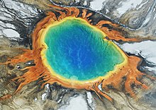
For much of the 20th century, prokaryotes were regarded as a single group of organisms and classified based on their biochemistry, morphology and metabolism. Microbiologists tried to classify microorganisms based on the structures of their cell walls, their shapes, and the substances they consume.[10] In 1965, Emile Zuckerkandl and Linus Pauling[11] instead proposed using the sequences of the genes in different prokaryotes to work out how they are related to each other. This phylogenetic approach is the main method used today.[12]
Archaea were first classified separately from bacteria in 1977 by Carl Woese and George E. Fox, based on their ribosomal RNA (rRNA) genes.[13] (At that time only the methanogens were known). They called these groups the Urkingdoms of Archaebacteria and Eubacteria, though other researchers treated them as kingdoms or subkingdoms. Woese and Fox gave the first evidence for Archaebacteria as a separate "line of descent": 1. lack of peptidoglycan in their cell walls, 2. two unusual coenzymes, 3. results of 16S ribosomal RNA gene sequencing. To emphasize this difference, Woese, Otto Kandler and Mark Wheelis later proposed reclassifying organisms into three natural domains known as the three-domain system: the Eukarya, the Bacteria and the Archaea,[1] in what is now known as the Woesian Revolution.[14]
The word archaea comes from the Ancient Greek ἀρχαῖα, meaning "ancient things",[15] as the first representatives of the domain Archaea were methanogens and it was assumed that their metabolism reflected Earth's primitive atmosphere and the organisms' antiquity, but as new habitats were studied, more organisms were discovered. Extreme halophilic[16] and hyperthermophilic microbes[17] were also included in Archaea. For a long time, archaea were seen as extremophiles that exist only in extreme habitats such as hot springs and salt lakes, but by the end of the 20th century, archaea had been identified in non-extreme environments as well. Today, they are known to be a large and diverse group of organisms abundantly distributed throughout nature.[18] This new appreciation of the importance and ubiquity of archaea came from using polymerase chain reaction (PCR) to detect prokaryotes from environmental samples (such as water or soil) by multiplying their ribosomal genes. This allows the detection and identification of organisms that have not been cultured in the laboratory.[19][20]
Classification
[edit]
The classification of archaea, and of prokaryotes in general, is a rapidly moving and contentious field. Current classification systems aim to organize archaea into groups of organisms that share structural features and common ancestors.[21] These classifications rely heavily on the use of the sequence of ribosomal RNA genes to reveal relationships among organisms (molecular phylogenetics).[22] Most of the culturable and well-investigated species of archaea are members of two main phyla, the "Euryarchaeota" and the Thermoproteota (formerly Crenarchaeota). Other groups have been tentatively created, such as the peculiar species Nanoarchaeum equitans — discovered in 2003 and assigned its own phylum, the "Nanoarchaeota".[23] A new phylum "Korarchaeota" has also been proposed, containing a small group of unusual thermophilic species sharing features of both the main phyla, but most closely related to the Thermoproteota.[24][25] Other detected species of archaea are only distantly related to any of these groups, such as the Archaeal Richmond Mine acidophilic nanoorganisms (ARMAN, comprising Micrarchaeota and Parvarchaeota), which were discovered in 2006[26] and are some of the smallest organisms known.[27]
A superphylum – TACK – which includes the Thaumarchaeota (now Nitrososphaerota), "Aigarchaeota", Crenarchaeota (now Thermoproteota), and "Korarchaeota" was proposed in 2011 to be related to the origin of eukaryotes.[28] In 2017, the newly discovered and newly named Asgard superphylum was proposed to be more closely related to the original eukaryote and a sister group to TACK.[29]
In 2013, the superphylum DPANN was proposed to group "Nanoarchaeota", "Nanohaloarchaeota", Archaeal Richmond Mine acidophilic nanoorganisms (ARMAN, comprising "Micrarchaeota" and "Parvarchaeota"), and other similar archaea. This archaeal superphylum encompasses at least 10 different lineages and includes organisms with extremely small cell and genome sizes and limited metabolic capabilities. Therefore, DPANN may include members obligately dependent on symbiotic interactions, and may even include novel parasites. However, other phylogenetic analyses found that DPANN does not form a monophyletic group, and that the apparent grouping is caused by long branch attraction (LBA), suggesting that all these lineages belong to "Euryarchaeota".[30][2]
Cladogram
[edit]According to Tom A. Williams et al. 2017,[31] Castelle & Banfield (2018)[32] and GTDB release 08-RS214 (28 April 2023):[33][34][35]
| Tom A. Williams et al. 2017[31] and Castelle & Banfield 2018[32] | 08-RS214 (28 April 2023)[33][34][35] | |||||||||||||||||||||||||||||||||||||||||||||||||||||||||||||||||||||||||||||||||||||||||||||||||||||||||||||||||||||||||||||||||||||||||||||||||||||||||||||||||||||||||||||||||||||||||
|---|---|---|---|---|---|---|---|---|---|---|---|---|---|---|---|---|---|---|---|---|---|---|---|---|---|---|---|---|---|---|---|---|---|---|---|---|---|---|---|---|---|---|---|---|---|---|---|---|---|---|---|---|---|---|---|---|---|---|---|---|---|---|---|---|---|---|---|---|---|---|---|---|---|---|---|---|---|---|---|---|---|---|---|---|---|---|---|---|---|---|---|---|---|---|---|---|---|---|---|---|---|---|---|---|---|---|---|---|---|---|---|---|---|---|---|---|---|---|---|---|---|---|---|---|---|---|---|---|---|---|---|---|---|---|---|---|---|---|---|---|---|---|---|---|---|---|---|---|---|---|---|---|---|---|---|---|---|---|---|---|---|---|---|---|---|---|---|---|---|---|---|---|---|---|---|---|---|---|---|---|---|---|---|---|---|---|
|
|
Concept of species
[edit]The classification of archaea into species is also controversial. Ernst Mayr's species definition — a reproductively isolated group of interbreeding organisms — does not apply, as archaea reproduce only asexually.[37]
Archaea show high levels of horizontal gene transfer between lineages. Some researchers suggest that individuals can be grouped into species-like populations given highly similar genomes and infrequent gene transfer to/from cells with less-related genomes, as in the genus Ferroplasma.[38] On the other hand, studies in Halorubrum found significant genetic transfer to/from less-related populations, limiting the criterion's applicability.[39] Some researchers question whether such species designations have practical meaning.[40]
Current knowledge on genetic diversity in archaeans is fragmentary, so the total number of species cannot be estimated with any accuracy.[22] Estimates of the number of phyla range from 18 to 23, of which only 8 have representatives that have been cultured and studied directly. Many of these hypothesized groups are known from a single rRNA sequence, so the level of diversity remains obscure.[41] This situation is also seen in the Bacteria; many uncultured microbes present similar issues with characterization.[42]
Phyla
[edit]Valid phyla
[edit]The following phyla have been validly published according to the Bacteriological Code:[43][44]
Provisional phyla
[edit]The following phyla have been proposed, but have not been validly published according to the Bacteriological Code (including those that have candidatus status):
- "Candidatus Aenigmarchaeota"
- "Candidatus Aigarchaeota"
- "Candidatus Altiarchaeota"
- "Candidatus Asgardaeota"
- "Candidatus Bathyarchaeota"
- "Candidatus Brockarchaeota"
- "Candidatus Diapherotrites"
- "Euryarchaeota"
- "Candidatus Geoarchaeota"
- "Candidatus Hadarchaeota"
- "Candidatus Hadesarchaeota"
- "Candidatus Halobacterota"
- "Candidatus Heimdallarchaeota"
- "Candidatus Helarchaeota"
- "Candidatus Huberarchaeota"
- "Candidatus Hydrothermarchaeota"
- "Candidatus Korarchaeota"
- "Candidatus Lokiarchaeia"
- "Candidatus Lokiarchaeota"
- "Candidatus Mamarchaeota"
- "Candidatus Marsarchaeota"
- "Candidatus Micrarchaeota"
- "Candidatus Nanoarchaeota"
- "Candidatus Nanohaloarchaeota"
- "Candidatus Nezhaarchaeota"
- "Candidatus Odinarchaeota"
- "Candidatus Pacearchaeota"
- "Candidatus Parvarchaeota"
- "Candidatus Thermoplasmatota"
- "Candidatus Thorarchaeota"
- "Candidatus Undinarchaeota"
- "Candidatus Verstraetearchaeota"
- "Candidatus Woesearchaeota"
Origin and evolution
[edit]The age of the Earth is about 4.54 billion years.[45][46][47] Scientific evidence suggests that life began on Earth at least 3.5 billion years ago.[48][49] The earliest evidence for life on Earth is graphite found to be biogenic in 3.7-billion-year-old metasedimentary rocks discovered in Western Greenland[50] and microbial mat fossils found in 3.48-billion-year-old sandstone discovered in Western Australia.[51][52] In 2015, possible remains of biotic matter were found in 4.1-billion-year-old rocks in Western Australia.[53][54]
Although probable prokaryotic cell fossils date to almost 3.5 billion years ago, most prokaryotes do not have distinctive morphologies, and fossil shapes cannot be used to identify them as archaea.[55] Instead, chemical fossils of unique lipids are more informative because such compounds do not occur in other organisms.[56] Some publications suggest that archaeal or eukaryotic lipid remains are present in shales dating from 2.7 billion years ago,[57] though such data have since been questioned.[58] These lipids have also been detected in even older rocks from west Greenland. The oldest such traces come from the Isua district, which includes Earth's oldest known sediments, formed 3.8 billion years ago.[59] The archaeal lineage may be the most ancient that exists on Earth.[60]
Woese argued that the Bacteria, Archaea, and Eukaryotes represent separate lines of descent that diverged early on from an ancestral colony of organisms.[61][62] One possibility[62][63] is that this occurred before the evolution of cells, when the lack of a typical cell membrane allowed unrestricted lateral gene transfer, and that the common ancestors of the three domains arose by fixation of specific subsets of genes.[62][63] It is possible that the last common ancestor of bacteria and archaea was a thermophile, which raises the possibility that lower temperatures are "extreme environments" for archaea, and organisms that live in cooler environments appeared only later.[64] Since archaea and bacteria are no more related to each other than they are to eukaryotes, the term prokaryote may suggest a false similarity between them.[65] However, structural and functional similarities between lineages often occur because of shared ancestral traits or evolutionary convergence. These similarities are known as a grade, and prokaryotes are best thought of as a grade of life, characterized by such features as an absence of membrane-bound organelles.
Comparison with other domains
[edit]The following table compares some major characteristics of the three domains, to illustrate their similarities and differences.[66]
| Property | Archaea | Bacteria | Eukaryota |
|---|---|---|---|
| Cell membrane | Ether-linked lipids | Ester-linked lipids | Ester-linked lipids |
| Cell wall | Glycoprotein, or S-layer; rarely pseudopeptidoglycan | Peptidoglycan, S-layer, or no cell wall | Various structures |
| Gene structure | Circular chromosomes, similar translation and transcription to Eukaryota | Circular chromosomes, unique translation and transcription | Multiple, linear chromosomes, but translation and transcription similar to Archaea |
| Internal cell structure | No membrane-bound organelles (?[67]) or nucleus | No membrane-bound organelles or nucleus | Membrane-bound organelles and nucleus |
| Metabolism[68] | Various, including diazotrophy, with methanogenesis unique to Archaea | Various, including photosynthesis, aerobic and anaerobic respiration, fermentation, diazotrophy, and autotrophy | Photosynthesis, cellular respiration, and fermentation; no diazotrophy |
| Reproduction | Asexual reproduction, horizontal gene transfer | Asexual reproduction, horizontal gene transfer | Sexual and asexual reproduction |
| Protein synthesis initiation | Methionine | Formylmethionine | Methionine |
| RNA polymerase | One | One | Many |
| EF-2/EF-G | Sensitive to diphtheria toxin | Resistant to diphtheria toxin | Sensitive to diphtheria toxin |
Archaea were split off as a third domain because of the large differences in their ribosomal RNA structure. The particular molecule 16S rRNA is key to the production of proteins in all organisms. Because this function is so central to life, organisms with mutations in their 16S rRNA are unlikely to survive, leading to great (but not absolute) stability in the structure of this polynucleotide over generations. 16S rRNA is large enough to show organism-specific variations, but still small enough to be compared quickly. In 1977, Carl Woese, a microbiologist studying the genetic sequences of organisms, developed a new comparison method that involved splitting the RNA into fragments that could be sorted and compared with other fragments from other organisms.[13] The more similar the patterns between species, the more closely they are related.[69]
Woese used his new rRNA comparison method to categorize and contrast different organisms. He compared a variety of species and happened upon a group of methanogens with rRNA vastly different from any known prokaryotes or eukaryotes.[13] These methanogens were much more similar to each other than to other organisms, leading Woese to propose the new domain of Archaea.[13] His experiments showed that the archaea were genetically more similar to eukaryotes than prokaryotes, even though they were more similar to prokaryotes in structure.[70] This led to the conclusion that Archaea and Eukarya shared a common ancestor more recent than Eukarya and Bacteria.[70] The development of the nucleus occurred after the split between Bacteria and this common ancestor.[70][1]
One property unique to archaea is the abundant use of ether-linked lipids in their cell membranes. Ether linkages are more chemically stable than the ester linkages found in bacteria and eukarya, which may be a contributing factor to the ability of many archaea to survive in extreme environments that place heavy stress on cell membranes, such as extreme heat and salinity. Comparative analysis of archaeal genomes has also identified several molecular conserved signature indels and signature proteins uniquely present in either all archaea or different main groups within archaea.[71][72][73] Another unique feature of archaea, found in no other organisms, is methanogenesis (the metabolic production of methane). Methanogenic archaea play a pivotal role in ecosystems with organisms that derive energy from oxidation of methane, many of which are bacteria, as they are often a major source of methane in such environments and can play a role as primary producers. Methanogens also play a critical role in the carbon cycle, breaking down organic carbon into methane, which is also a major greenhouse gas.[74]
This difference in the biochemical structure of Bacteria and Archaea has been explained by researchers through evolutionary processes. It is theorized that both domains originated at deep sea alkaline hydrothermal vents. At least twice, microbes evolved lipid biosynthesis and cell wall biochemistry. It has been suggested that the last universal common ancestor was a non-free-living organism.[75] It may have had a permeable membrane composed of bacterial simple chain amphiphiles (fatty acids), including archaeal simple chain amphiphiles (isoprenoids). These stabilize fatty acid membranes in seawater; this property may have driven the divergence of bacterial and archaeal membranes, "with the later biosynthesis of phospholipids giving rise to the unique G1P and G3P headgroups of archaea and bacteria respectively. If so, the properties conferred by membrane isoprenoids place the lipid divide as early as the origin of life".[76]
Relationship to bacteria
[edit]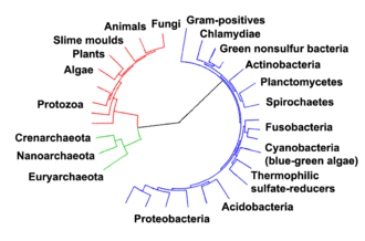
The relationships among the three domains are of central importance for understanding the origin of life. Most of the metabolic pathways, which are the object of the majority of an organism's genes, are common between Archaea and Bacteria, while most genes involved in genome expression are common between Archaea and Eukarya.[78] Within prokaryotes, archaeal cell structure is most similar to that of gram-positive bacteria, largely because both have a single lipid bilayer[79] and usually contain a thick sacculus (exoskeleton) of varying chemical composition.[80] In some phylogenetic trees based upon different gene/protein sequences of prokaryotic homologs, the archaeal homologs are more closely related to those of gram-positive bacteria.[79] Archaea and gram-positive bacteria also share conserved indels in a number of important proteins, such as Hsp70 and glutamine synthetase I;[79][81] but the phylogeny of these genes was interpreted to reveal interdomain gene transfer,[82][83] and might not reflect the organismal relationship(s).[84]
It has been proposed that the archaea evolved from gram-positive bacteria in response to antibiotic selection pressure.[79][81][85] This is suggested by the observation that archaea are resistant to a wide variety of antibiotics that are produced primarily by gram-positive bacteria,[79][81] and that these antibiotics act primarily on the genes that distinguish archaea from bacteria. The proposal is that the selective pressure towards resistance generated by the gram-positive antibiotics was eventually sufficient to cause extensive changes in many of the antibiotics' target genes, and that these strains represented the common ancestors of present-day Archaea.[85] The evolution of Archaea in response to antibiotic selection, or any other competitive selective pressure, could also explain their adaptation to extreme environments (such as high temperature or acidity) as the result of a search for unoccupied niches to escape from antibiotic-producing organisms;[85][86] Cavalier-Smith has made a similar suggestion.[87] This proposal is also supported by other work investigating protein structural relationships[88] and studies that suggest that gram-positive bacteria may constitute the earliest branching lineages within the prokaryotes.[89]
Relation to eukaryotes
[edit]
The evolutionary relationship between archaea and eukaryotes remains unclear. Aside from the similarities in cell structure and function that are discussed below, many genetic trees group the two.[91]
Complicating factors include claims that the relationship between eukaryotes and the archaeal phylum Thermoproteota is closer than the relationship between the "Euryarchaeota" and the phylum Thermoproteota[92] and the presence of archaea-like genes in certain bacteria, such as Thermotoga maritima, from horizontal gene transfer.[93] The standard hypothesis states that the ancestor of the eukaryotes diverged early from the Archaea,[94][95] and that eukaryotes arose through symbiogenesis, the fusion of an archaean and a eubacterium, which formed the mitochondria; this hypothesis explains the genetic similarities between the groups.[90] The eocyte hypothesis instead posits that Eukaryota emerged relatively late from the Archaea.[96]
A lineage of archaea discovered in 2015, Lokiarchaeum (of the proposed new phylum "Lokiarchaeota"), named for a hydrothermal vent called Loki's Castle in the Arctic Ocean, was found to be the most closely related to eukaryotes known at that time. It has been called a transitional organism between prokaryotes and eukaryotes.[97][98]
Several sister phyla of "Lokiarchaeota" have since been found ("Thorarchaeota", "Odinarchaeota", "Heimdallarchaeota"), all together comprising a newly proposed supergroup Asgard.[29][3][99]
Details of the relation of Asgard members and eukaryotes are still under consideration,[100] although, in January 2020, scientists reported that Candidatus Prometheoarchaeum syntrophicum, a type of Asgard archaea, may be a possible link between simple prokaryotic and complex eukaryotic microorganisms about two billion years ago.[101][102][103]
Morphology
[edit]Individual archaea range from 0.1 micrometers (μm) to over 15 μm in diameter, and occur in various shapes, commonly as spheres, rods, spirals or plates.[104] Other morphologies in the Thermoproteota include irregularly shaped lobed cells in Sulfolobus, needle-like filaments that are less than half a micrometer in diameter in Thermofilum, and almost perfectly rectangular rods in Thermoproteus and Pyrobaculum.[105] Archaea in the genus Haloquadratum such as Haloquadratum walsbyi are flat, square specimens that live in hypersaline pools.[106] These unusual shapes are probably maintained by both their cell walls and a prokaryotic cytoskeleton. Proteins related to the cytoskeleton components of other organisms exist in archaea,[107] and filaments form within their cells,[108] but in contrast with other organisms, these cellular structures are poorly understood.[109] In Thermoplasma and Ferroplasma the lack of a cell wall means that the cells have irregular shapes, and can resemble amoebae.[110]
Some species form aggregates or filaments of cells up to 200 μm long.[104] These organisms can be prominent in biofilms.[111] Notably, aggregates of Thermococcus coalescens cells fuse together in culture, forming single giant cells.[112] Archaea in the genus Pyrodictium produce an elaborate multicell colony involving arrays of long, thin hollow tubes called cannulae that stick out from the cells' surfaces and connect them into a dense bush-like agglomeration.[113] The function of these cannulae is not settled, but they may allow communication or nutrient exchange with neighbors.[114] Multi-species colonies exist, such as the "string-of-pearls" community that was discovered in 2001 in a German swamp. Round whitish colonies of a novel Euryarchaeota species are spaced along thin filaments that can range up to 15 centimetres (5.9 in) long; these filaments are made of a particular bacteria species.[115]
Structure, composition development, and operation
[edit]Archaea and bacteria have generally similar cell structure, but cell composition and organization set the archaea apart. Like bacteria, archaea lack interior membranes and organelles.[65] Like bacteria, the cell membranes of archaea are usually bounded by a cell wall and they swim using one or more flagella.[116] Structurally, archaea are most similar to gram-positive bacteria. Most have a single plasma membrane and cell wall, and lack a periplasmic space; the exception to this general rule is Ignicoccus, which possess a particularly large periplasm that contains membrane-bound vesicles and is enclosed by an outer membrane.[117]
Cell wall and archaella
[edit]Most archaea (but not Thermoplasma and Ferroplasma) possess a cell wall.[110] In most archaea, the wall is assembled from surface-layer proteins, which form an S-layer.[118] An S-layer is a rigid array of protein molecules that cover the outside of the cell (like chain mail).[119] This layer provides both chemical and physical protection, and can prevent macromolecules from contacting the cell membrane.[120] Unlike bacteria, archaea lack peptidoglycan in their cell walls.[121] Methanobacteriales do have cell walls containing pseudopeptidoglycan, which resembles eubacterial peptidoglycan in morphology, function, and physical structure, but pseudopeptidoglycan is distinct in chemical structure; it lacks D-amino acids and N-acetylmuramic acid, substituting the latter with N-Acetyltalosaminuronic acid.[120]
Archaeal flagella are known as archaella, that operate like bacterial flagella – their long stalks are driven by rotatory motors at the base. These motors are powered by a proton gradient across the membrane, but archaella are notably different in composition and development.[116] The two types of flagella evolved from different ancestors. The bacterial flagellum shares a common ancestor with the type III secretion system,[122][123] while archaeal flagella appear to have evolved from bacterial type IV pili.[124] In contrast with the bacterial flagellum, which is hollow and assembled by subunits moving up the central pore to the tip of the flagella, archaeal flagella are synthesized by adding subunits at the base.[125]
Membranes
[edit]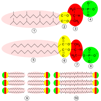
Archaeal membranes are made of molecules that are distinctly different from those in all other life forms, showing that archaea are related only distantly to bacteria and eukaryotes.[126] In all organisms, cell membranes are made of molecules known as phospholipids. These molecules possess both a polar part that dissolves in water (the phosphate "head"), and a "greasy" non-polar part that does not (the lipid tail). These dissimilar parts are connected by a glycerol moiety. In water, phospholipids cluster, with the heads facing the water and the tails facing away from it. The major structure in cell membranes is a double layer of these phospholipids, which is called a lipid bilayer.[127]
The phospholipids of archaea are unusual in four ways:
- They have membranes composed of glycerol-ether lipids, whereas bacteria and eukaryotes have membranes composed mainly of glycerol-ester lipids.[128] The difference is the type of bond that joins the lipids to the glycerol moiety; the two types are shown in yellow in the figure at the right. In ester lipids this is an ester bond, whereas in ether lipids this is an ether bond.[129]
- The stereochemistry of the archaeal glycerol moiety is the mirror image of that found in other organisms. The glycerol moiety can occur in two forms that are mirror images of one another, called enantiomers. Just as a right hand does not fit easily into a left-handed glove, enantiomers of one type generally cannot be used or made by enzymes adapted for the other. The archaeal phospholipids are built on a backbone of sn-glycerol-1-phosphate, which is an enantiomer of sn-glycerol-3-phosphate, the phospholipid backbone found in bacteria and eukaryotes. This suggests that archaea use entirely different enzymes for synthesizing phospholipids as compared to bacteria and eukaryotes. Such enzymes developed very early in life's history, indicating an early split from the other two domains.[126]
- Archaeal lipid tails differ from those of other organisms in that they are based upon long isoprenoid chains with multiple side-branches, sometimes with cyclopropane or cyclohexane rings.[130] By contrast, the fatty acids in the membranes of other organisms have straight chains without side branches or rings. Although isoprenoids play an important role in the biochemistry of many organisms, only the archaea use them to make phospholipids. These branched chains may help prevent archaeal membranes from leaking at high temperatures.[131]
- In some archaea, the lipid bilayer is replaced by a monolayer. In effect, the archaea fuse the tails of two phospholipid molecules into a single molecule with two polar heads (a bolaamphiphile); this fusion may make their membranes more rigid and better able to resist harsh environments.[132] For example, the lipids in Ferroplasma are of this type, which is thought to aid this organism's survival in its highly acidic habitat.[133]
Metabolism
[edit]Archaea exhibit a great variety of chemical reactions in their metabolism and use many sources of energy. These reactions are classified into nutritional groups, depending on energy and carbon sources. Some archaea obtain energy from inorganic compounds such as sulfur or ammonia (they are chemotrophs). These include nitrifiers, methanogens and anaerobic methane oxidisers.[134] In these reactions one compound passes electrons to another (in a redox reaction), releasing energy to fuel the cell's activities. One compound acts as an electron donor and one as an electron acceptor. The energy released is used to generate adenosine triphosphate (ATP) through chemiosmosis, the same basic process that happens in the mitochondrion of eukaryotic cells.[135]
Other groups of archaea use sunlight as a source of energy (they are phototrophs), but oxygen–generating photosynthesis does not occur in any of these organisms.[135] Many basic metabolic pathways are shared among all forms of life; for example, archaea use a modified form of glycolysis (the Entner–Doudoroff pathway) and either a complete or partial citric acid cycle.[136] These similarities to other organisms probably reflect both early origins in the history of life and their high level of efficiency.[137]
| Nutritional type | Source of energy | Source of carbon | Examples |
|---|---|---|---|
| Phototrophs | Sunlight | Organic compounds | Halobacterium |
| Lithotrophs | Inorganic compounds | Organic compounds or carbon fixation | Ferroglobus, Methanobacteria or Pyrolobus |
| Organotrophs | Organic compounds | Organic compounds or carbon fixation | Pyrococcus, Sulfolobus or Methanosarcinales |
Some Euryarchaeota are methanogens (archaea that produce methane as a result of metabolism) living in anaerobic environments, such as swamps. This form of metabolism evolved early, and it is even possible that the first free-living organism was a methanogen.[138] A common reaction involves the use of carbon dioxide as an electron acceptor to oxidize hydrogen. Methanogenesis involves a range of coenzymes that are unique to these archaea, such as coenzyme M and methanofuran.[139] Other organic compounds such as alcohols, acetic acid or formic acid are used as alternative electron acceptors by methanogens. These reactions are common in gut-dwelling archaea. Acetic acid is also broken down into methane and carbon dioxide directly, by acetotrophic archaea. These acetotrophs are archaea in the order Methanosarcinales, and are a major part of the communities of microorganisms that produce biogas.[140]
Other archaea use CO
2 in the atmosphere as a source of carbon, in a process called carbon fixation (they are autotrophs). This process involves either a highly modified form of the Calvin cycle[142] or another metabolic pathway called the 3-hydroxypropionate/ 4-hydroxybutyrate cycle.[143] The Thermoproteota also use the reverse Krebs cycle while the "Euryarchaeota" also use the reductive acetyl-CoA pathway.[144] Carbon fixation is powered by inorganic energy sources. No known archaea carry out photosynthesis[145] (Halobacterium is the only known phototroph archeon but it uses an alternative process to photosynthesis). Archaeal energy sources are extremely diverse, and range from the oxidation of ammonia by the Nitrosopumilales[146][147] to the oxidation of hydrogen sulfide or elemental sulfur by species of Sulfolobus, using either oxygen or metal ions as electron acceptors.[135]
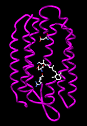
Phototrophic archaea use light to produce chemical energy in the form of ATP. In the Halobacteria, light-activated ion pumps like bacteriorhodopsin and halorhodopsin generate ion gradients by pumping ions out of and into the cell across the plasma membrane. The energy stored in these electrochemical gradients is then converted into ATP by ATP synthase.[104] This process is a form of photophosphorylation. The ability of these light-driven pumps to move ions across membranes depends on light-driven changes in the structure of a retinol cofactor buried in the center of the protein.[148]
Genetics
[edit]Archaea usually have a single circular chromosome,[149] but many euryarchaea have been shown to bear multiple copies of this chromosome.[150] The largest known archaeal genome as of 2002 was 5,751,492 base pairs in Methanosarcina acetivorans.[151] The tiny 490,885 base-pair genome of Nanoarchaeum equitans is one-tenth of this size and the smallest archaeal genome known; it is estimated to contain only 537 protein-encoding genes.[152] Smaller independent pieces of DNA, called plasmids, are also found in archaea. Plasmids may be transferred between cells by physical contact, in a process that may be similar to bacterial conjugation.[153][154]
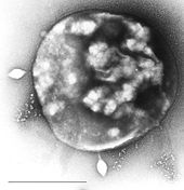
Archaea are genetically distinct from bacteria and eukaryotes, with up to 15% of the proteins encoded by any one archaeal genome being unique to the domain, although most of these unique genes have no known function.[156] Of the remainder of the unique proteins that have an identified function, most belong to the Euryarchaeota and are involved in methanogenesis. The proteins that archaea, bacteria and eukaryotes share form a common core of cell function, relating mostly to transcription, translation, and nucleotide metabolism.[157] Other characteristic archaeal features are the organization of genes of related function – such as enzymes that catalyze steps in the same metabolic pathway into novel operons, and large differences in tRNA genes and their aminoacyl tRNA synthetases.[157]
Transcription in archaea more closely resembles eukaryotic than bacterial transcription, with the archaeal RNA polymerase being very close to its equivalent in eukaryotes,[149] while archaeal translation shows signs of both bacterial and eukaryotic equivalents.[158] Although archaea have only one type of RNA polymerase, its structure and function in transcription seems to be close to that of the eukaryotic RNA polymerase II, with similar protein assemblies (the general transcription factors) directing the binding of the RNA polymerase to a gene's promoter,[159] but other archaeal transcription factors are closer to those found in bacteria.[160] Post-transcriptional modification is simpler than in eukaryotes, since most archaeal genes lack introns, although there are many introns in their transfer RNA and ribosomal RNA genes,[161] and introns may occur in a few protein-encoding genes.[162][163]
Gene transfer and genetic exchange
[edit]Haloferax volcanii, an extreme halophilic archaeon, forms cytoplasmic bridges between cells that appear to be used for transfer of DNA from one cell to another in either direction.[164]
When the hyperthermophilic archaea Sulfolobus solfataricus[165] and Sulfolobus acidocaldarius[166] are exposed to DNA-damaging UV irradiation or to the agents bleomycin or mitomycin C, species-specific cellular aggregation is induced. Aggregation in S. solfataricus could not be induced by other physical stressors, such as pH or temperature shift,[165] suggesting that aggregation is induced specifically by DNA damage. Ajon et al.[166] showed that UV-induced cellular aggregation mediates chromosomal marker exchange with high frequency in S. acidocaldarius. Recombination rates exceeded those of uninduced cultures by up to three orders of magnitude. Frols et al.[165][167] and Ajon et al.[166] hypothesized that cellular aggregation enhances species-specific DNA transfer between Sulfolobus cells in order to provide increased repair of damaged DNA by means of homologous recombination. This response may be a primitive form of sexual interaction similar to the more well-studied bacterial transformation systems that are also associated with species-specific DNA transfer between cells leading to homologous recombinational repair of DNA damage.[168]
Archaeal viruses
[edit]Archaea are the target of a number of viruses in a diverse virosphere distinct from bacterial and eukaryotic viruses. They have been organized into 15–18 DNA-based families so far, but multiple species remain un-isolated and await classification.[169][170][171] These families can be informally divided into two groups: archaea-specific and cosmopolitan. Archaeal-specific viruses target only archaean species and currently include 12 families. Numerous unique, previously unidentified viral structures have been observed in this group, including: bottle-shaped, spindle-shaped, coil-shaped, and droplet-shaped viruses.[170] While the reproductive cycles and genomic mechanisms of archaea-specific species may be similar to other viruses, they bear unique characteristics that were specifically developed due to the morphology of host cells they infect.[169] Their virus release mechanisms differ from that of other phages. Bacteriophages generally undergo either lytic pathways, lysogenic pathways, or (rarely) a mix of the two.[172] Most archaea-specific viral strains maintain a stable, somewhat lysogenic, relationship with their hosts – appearing as a chronic infection. This involves the gradual, and continuous, production and release of virions without killing the host cell.[173] Prangishyili (2013) noted that it has been hypothesized that tailed archaeal phages originated from bacteriophages capable of infecting haloarchaeal species. If the hypothesis is correct, it can be concluded that other double-stranded DNA viruses that make up the rest of the archaea-specific group are their own unique group in the global viral community. Krupovic et al. (2018) states that the high levels of horizontal gene transfer, rapid mutation rates in viral genomes, and lack of universal gene sequences have led researchers to perceive the evolutionary pathway of archaeal viruses as a network. The lack of similarities among phylogenetic markers in this network and the global virosphere, as well as external linkages to non-viral elements, may suggest that some species of archaea specific viruses evolved from non-viral mobile genetic elements (MGE).[170]
These viruses have been studied in most detail in thermophilics, particularly the orders Sulfolobales and Thermoproteales.[174] Two groups of single-stranded DNA viruses that infect archaea have been recently isolated. One group is exemplified by the Halorubrum pleomorphic virus 1 (Pleolipoviridae) infecting halophilic archaea,[175] and the other one by the Aeropyrum coil-shaped virus (Spiraviridae) infecting a hyperthermophilic (optimal growth at 90–95 °C) host.[176] Notably, the latter virus has the largest currently reported ssDNA genome. Defenses against these viruses may involve RNA interference from repetitive DNA sequences that are related to the genes of the viruses.[177][178]
Reproduction
[edit]Archaea reproduce asexually by binary or multiple fission, fragmentation, or budding; mitosis and meiosis do not occur, so if a species of archaea exists in more than one form, all have the same genetic material.[104] Cell division is controlled in a cell cycle; after the cell's chromosome is replicated and the two daughter chromosomes separate, the cell divides.[179] In the genus Sulfolobus, the cycle has characteristics that are similar to both bacterial and eukaryotic systems. The chromosomes replicate from multiple starting points (origins of replication) using DNA polymerases that resemble the equivalent eukaryotic enzymes.[180]
In Euryarchaeota the cell division protein FtsZ, which forms a contracting ring around the cell, and the components of the septum that is constructed across the center of the cell, are similar to their bacterial equivalents.[179] In cren-[181][182] and thaumarchaea,[183] the cell division machinery Cdv fulfills a similar role. This machinery is related to the eukaryotic ESCRT-III machinery which, while best known for its role in cell sorting, also has been seen to fulfill a role in separation between divided cell, suggesting an ancestral role in cell division.[184]
Both bacteria and eukaryotes, but not archaea, make spores.[185] Some species of Haloarchaea undergo phenotypic switching and grow as several different cell types, including thick-walled structures that are resistant to osmotic shock and allow the archaea to survive in water at low salt concentrations, but these are not reproductive structures and may instead help them reach new habitats.[186]
Behavior
[edit]Communication
[edit]Quorum sensing was originally thought to not exist in Archaea, but recent studies have shown evidence of some species being able to perform cross-talk through quorum sensing. Other studies have shown syntrophic interactions between archaea and bacteria during biofilm growth. Although research is limited in archaeal quorum sensing, some studies have uncovered LuxR proteins in archaeal species, displaying similarities with bacteria LuxR, and ultimately allowing for the detection of small molecules that are used in high density communication. Similarly to bacteria, Archaea LuxR solos have shown to bind to AHLs (lactones) and non-AHLs ligans, which is a large part in performing intraspecies, interspecies, and interkingdom communication through quorum sensing.[187]
Ecology
[edit]Habitats
[edit]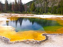
Archaea exist in a broad range of habitats, and are now recognized as a major part of global ecosystems,[18] and may represent about 20% of microbial cells in the oceans.[188] However, the first-discovered archaeans were extremophiles.[134] Indeed, some archaea survive high temperatures, often above 100 °C (212 °F), as found in geysers, black smokers, and oil wells. Other common habitats include very cold habitats and highly saline, acidic, or alkaline water, but archaea include mesophiles that grow in mild conditions, in swamps and marshland, sewage, the oceans, the intestinal tract of animals, and soils.[7][18] Similar to PGPR, Archaea are now considered as a source of plant growth promotion as well.[7]
Extremophile archaea are members of four main physiological groups. These are the halophiles, thermophiles, alkaliphiles, and acidophiles.[189] These groups are not comprehensive or phylum-specific, nor are they mutually exclusive, since some archaea belong to several groups. Nonetheless, they are a useful starting point for classification.[190]
Halophiles, including the genus Halobacterium, live in extremely saline environments such as salt lakes and outnumber their bacterial counterparts at salinities greater than 20–25%.[134] Thermophiles grow best at temperatures above 45 °C (113 °F), in places such as hot springs; hyperthermophilic archaea grow optimally at temperatures greater than 80 °C (176 °F).[191] The archaeal Methanopyrus kandleri Strain 116 can even reproduce at 122 °C (252 °F), the highest recorded temperature of any organism.[192]
Other archaea exist in very acidic or alkaline conditions.[189] For example, one of the most extreme archaean acidophiles is Picrophilus torridus, which grows at pH 0, which is equivalent to thriving in 1.2 molar sulfuric acid.[193]
This resistance to extreme environments has made archaea the focus of speculation about the possible properties of extraterrestrial life.[194] Some extremophile habitats are not dissimilar to those on Mars,[195] leading to the suggestion that viable microbes could be transferred between planets in meteorites.[196]
Recently, several studies have shown that archaea exist not only in mesophilic and thermophilic environments but are also present, sometimes in high numbers, at low temperatures as well. For example, archaea are common in cold oceanic environments such as polar seas.[197] Even more significant are the large numbers of archaea found throughout the world's oceans in non-extreme habitats among the plankton community (as part of the picoplankton).[198] Although these archaea can be present in extremely high numbers (up to 40% of the microbial biomass), almost none of these species have been isolated and studied in pure culture.[199] Consequently, our understanding of the role of archaea in ocean ecology is rudimentary, so their full influence on global biogeochemical cycles remains largely unexplored.[200] Some marine Thermoproteota are capable of nitrification, suggesting these organisms may affect the oceanic nitrogen cycle,[146] although these oceanic Thermoproteota may also use other sources of energy.[201]
Vast numbers of archaea are also found in the sediments that cover the sea floor, with these organisms making up the majority of living cells at depths over 1 meter below the ocean bottom.[202][203] It has been demonstrated that in all oceanic surface sediments (from 1000- to 10,000-m water depth), the impact of viral infection is higher on archaea than on bacteria and virus-induced lysis of archaea accounts for up to one-third of the total microbial biomass killed, resulting in the release of ~0.3 to 0.5 gigatons of carbon per year globally.[204]
Role in chemical cycling
[edit]Archaea recycle elements such as carbon, nitrogen, and sulfur through their various habitats.[205] Archaea carry out many steps in the nitrogen cycle. This includes both reactions that remove nitrogen from ecosystems (such as nitrate-based respiration and denitrification) as well as processes that introduce nitrogen (such as nitrate assimilation and nitrogen fixation).[206][207] Researchers recently discovered archaeal involvement in ammonia oxidation reactions. These reactions are particularly important in the oceans.[147][208] The archaea also appear crucial for ammonia oxidation in soils. They produce nitrite, which other microbes then oxidize to nitrate. Plants and other organisms consume the latter.[209]
In the sulfur cycle, archaea that grow by oxidizing sulfur compounds release this element from rocks, making it available to other organisms, but the archaea that do this, such as Sulfolobus, produce sulfuric acid as a waste product, and the growth of these organisms in abandoned mines can contribute to acid mine drainage and other environmental damage.[210]
In the carbon cycle, methanogen archaea remove hydrogen and play an important role in the decay of organic matter by the populations of microorganisms that act as decomposers in anaerobic ecosystems, such as sediments, marshes, and sewage-treatment works.[211]
Interactions with other organisms
[edit]
The well-characterized interactions between archaea and other organisms are either mutual or commensal. There are no clear examples of known archaeal pathogens or parasites,[212][213] but some species of methanogens have been suggested to be involved in infections in the mouth,[214][215] and Nanoarchaeum equitans may be a parasite of another species of archaea, since it only survives and reproduces within the cells of the Crenarchaeon Ignicoccus hospitalis,[152] and appears to offer no benefit to its host.[216]
Mutualism
[edit]Mutualism is an interaction between individuals of different species that results in positive (beneficial) effects on per capita reproduction and/or survival of the interacting populations. One well-understood example of mutualism is the interaction between protozoa and methanogenic archaea in the digestive tracts of animals that digest cellulose, such as ruminants and termites.[217] In these anaerobic environments, protozoa break down plant cellulose to obtain energy. This process releases hydrogen as a waste product, but high levels of hydrogen reduce energy production. When methanogens convert hydrogen to methane, protozoa benefit from more energy.[218]
In anaerobic protozoa, such as Plagiopyla frontata, Trimyema, Heterometopus and Metopus contortus, archaea reside inside the protozoa and consume hydrogen produced in their hydrogenosomes.[219][220][221][222][223] Archaea associate with larger organisms, too. For example, the marine archaean Cenarchaeum symbiosum is an endosymbiont of the sponge Axinella mexicana.[224]
Commensalism
[edit]Some archaea are commensals, benefiting from an association without helping or harming the other organism. For example, the methanogen Methanobrevibacter smithii is by far the most common archaean in the human flora, making up about one in ten of the prokaryotes in the human gut.[225] In termites and in humans, these methanogens may in fact be mutualists, interacting with other microbes in the gut to aid digestion.[226] Archaean communities associate with a range of other organisms, such as on the surface of corals,[227] and in the region of soil that surrounds plant roots (the rhizosphere).[228][229]
Parasitism
[edit]Although Archaea do not have a historical reputation of being pathogens, Archaea are often found with similar genomes to more common pathogen like E. coli,[230] showing metabolic links and evolutionary history with today's pathogens. Archaea have been inconsistently detected in clinical studies because of the lack of categorization of Archaea into more specific species.[231]
Significance in technology and industry
[edit]Extremophile archaea, particularly those resistant either to heat or to extremes of acidity and alkalinity, are a source of enzymes that function under these harsh conditions.[232][233] These enzymes have found many uses. For example, thermostable DNA polymerases, such as the Pfu DNA polymerase from Pyrococcus furiosus, revolutionized molecular biology by allowing the polymerase chain reaction to be used in research as a simple and rapid technique for cloning DNA. In industry, amylases, galactosidases and pullulanases in other species of Pyrococcus that function at over 100 °C (212 °F) allow food processing at high temperatures, such as the production of low lactose milk and whey.[234] Enzymes from these thermophilic archaea also tend to be very stable in organic solvents, allowing their use in environmentally friendly processes in green chemistry that synthesize organic compounds.[233] This stability makes them easier to use in structural biology. Consequently, the counterparts of bacterial or eukaryotic enzymes from extremophile archaea are often used in structural studies.[235]
In contrast with the range of applications of archaean enzymes, the use of the organisms themselves in biotechnology is less developed. Methanogenic archaea are a vital part of sewage treatment, since they are part of the community of microorganisms that carry out anaerobic digestion and produce biogas.[236] In mineral processing, acidophilic archaea display promise for the extraction of metals from ores, including gold, cobalt and copper.[237]
Archaea host a new class of potentially useful antibiotics. A few of these archaeocins have been characterized, but hundreds more are believed to exist, especially within Haloarchaea and Sulfolobus. These compounds differ in structure from bacterial antibiotics, so they may have novel modes of action. In addition, they may allow the creation of new selectable markers for use in archaeal molecular biology.[238]
See also
[edit]- Aerobic methane production
- Earliest known life forms
- List of Archaea genera
- List of sequenced archaeal genomes
- Nuclear localization sequence
- The Surprising Archaea (book)
- Towards a natural system of organisms: proposal for the domains Archaea, Bacteria, and Eucarya
- Unique properties of hyperthermophilic archaea
- Branching order of bacterial phyla (Genome Taxonomy Database, 2018)
References
[edit]- ^ Перейти обратно: а б с Вёзе Ч.Р., Кандлер О., Уилис М.Л. (июнь 1990 г.). «На пути к естественной системе организмов: предложение по доменам архей, бактерий и эукариев» . Труды Национальной академии наук Соединенных Штатов Америки . 87 (12): 4576–9. Бибкод : 1990PNAS...87.4576W . дои : 10.1073/pnas.87.12.4576 . ПМК 54159 . ПМИД 2112744 .
- ^ Перейти обратно: а б Петижан К., Дешам П., Лопес-Гарсиа П., Морейра Д. (декабрь 2014 г.). «Укоренение домена архей с помощью филогеномного анализа поддерживает основу нового царства Proteoarchaeota» . Геномная биология и эволюция . 7 (1): 191–204. дои : 10.1093/gbe/evu274 . ПМЦ 4316627 . ПМИД 25527841 .
- ^ Перейти обратно: а б «Страница таксономии NCBI архей» .
- ^ Пейс НР (май 2006 г.). «Время перемен» . Природа . 441 (7091): 289. Бибкод : 2006Natur.441..289P . дои : 10.1038/441289a . ПМИД 16710401 . S2CID 4431143 .
- ^ Стоекениус В. (октябрь 1981 г.). «Квадратная бактерия Уолсби: тонкая структура ортогонального прокариота» . Журнал бактериологии . 148 (1): 352–60. дои : 10.1128/JB.148.1.352-360.1981 . ПМК 216199 . ПМИД 7287626 .
- ^ «Основная биология архей» . Март 2018.
- ^ Перейти обратно: а б с д Чоу С., Падда КП, Пури А, Чанвей КП (сентябрь 2022 г.). «Архаичный подход к современной проблеме: эндофитные археи для устойчивого сельского хозяйства». Современная микробиология . 79 (11): 322. doi : 10.1007/s00284-022-03016-y . ПМИД 36125558 . S2CID 252376815 .
- ^ Банг С., Шмитц Р.А. (сентябрь 2015 г.). «Археи, связанные с человеческими поверхностями: нельзя недооценивать» . Обзоры микробиологии FEMS . 39 (5): 631–48. дои : 10.1093/femsre/fuv010 . ПМИД 25907112 .
- ^ Мойссль-Айхингер С., Паузан М., Таффнер Дж., Берг Г., Банг С., Шмитц Р.А. (январь 2018 г.). «Археи — интерактивные компоненты сложных микробиомов». Тенденции в микробиологии . 26 (1): 70–85. дои : 10.1016/j.tim.2017.07.004 . ПМИД 28826642 .
- ^ Стейли Дж.Т. (ноябрь 2006 г.). «Дилемма бактериального вида и геномно-филогенетическая концепция вида» . Философские труды Лондонского королевского общества. Серия Б, Биологические науки . 361 (1475): 1899–909. дои : 10.1098/rstb.2006.1914 . ПМЦ 1857736 . ПМИД 17062409 .
- ^ Цукеркандл Э., Полинг Л. (март 1965 г.). «Молекулы как документы эволюционной истории». Журнал теоретической биологии . 8 (2): 357–66. Бибкод : 1965JThBi...8..357Z . дои : 10.1016/0022-5193(65)90083-4 . ПМИД 5876245 .
- ^ Паркс Д.Х., Чувочина М., Уэйт Д.В., Ринке С., Скаршевски А., Шомей П.А., Гугенхольц П. (ноябрь 2018 г.). «Стандартизированная таксономия бактерий, основанная на филогении генома, существенно пересматривает древо жизни». Природная биотехнология . 36 (10): 996–1004. дои : 10.1038/nbt.4229 . ПМИД 30148503 . S2CID 52093100 .
- ^ Перейти обратно: а б с д Woese CR, Fox GE (ноябрь 1977 г.). «Филогенетическая структура прокариотического домена: первичные царства» . Труды Национальной академии наук Соединенных Штатов Америки . 74 (11): 5088–90. Бибкод : 1977PNAS...74.5088W . дои : 10.1073/pnas.74.11.5088 . ПМК 432104 . ПМИД 270744 .
- ^ Сапп Дж (2009). Новые основы эволюции: на древе жизни . Нью-Йорк: Издательство Оксфордского университета. ISBN 978-0-19-973438-2 .
- ^ «Архея» . Интернет-словарь Мерриам-Вебстера . 2008 год . Проверено 1 июля 2008 года .
- ^ Магрум Л.Дж., Луерсен К.Р., Вёзе Ч.Р. (май 1978 г.). «Являются ли крайние галофилы на самом деле «бактериями»?». Журнал молекулярной эволюции . 11 (1): 1–8. Бибкод : 1978JMolE..11....1M . дои : 10.1007/bf01768019 . ПМИД 660662 . S2CID 1291732 .
- ^ Стеттер КО (1996). «Гипертермофилы в истории жизни». Симпозиум Фонда Ciba . 202 : 1–10, обсуждение 11–8. ПМИД 9243007 .
- ^ Перейти обратно: а б с Делонг Э.Ф. (декабрь 1998 г.). «Все в меру: археи как «неэкстремофилы» ». Текущее мнение в области генетики и развития . 8 (6): 649–54. дои : 10.1016/S0959-437X(98)80032-4 . ПМИД 9914204 .
- ^ Терон Дж., Клоэте Т.Э. (2000). «Молекулярные методы определения микробного разнообразия и структуры сообществ в природных средах». Критические обзоры по микробиологии . 26 (1): 37–57. дои : 10.1080/10408410091154174 . ПМИД 10782339 . S2CID 5829573 .
- ^ Шмидт Т.М. (сентябрь 2006 г.). «Зрелость микробной экологии» (PDF) . Международная микробиология . 9 (3): 217–23. ПМИД 17061212 . Архивировано из оригинала (PDF) 11 сентября 2008 года.
- ^ Геверс Д., Давиндт П., Вандам П., Виллемс А., Ванканнейт М., Свингс Дж. и др. (ноябрь 2006 г.). «Ступеньки к новой таксономии прокариот» . Философские труды Лондонского королевского общества. Серия Б, Биологические науки . 361 (1475): 1911–16. дои : 10.1098/rstb.2006.1915 . ПМК 1764938 . ПМИД 17062410 .
- ^ Перейти обратно: а б Робертсон С.Э., Харрис Дж.К., Спир Дж.Р., Пейс Н.Р. (декабрь 2005 г.). «Филогенетическое разнообразие и экология природных архей». Современное мнение в микробиологии . 8 (6): 638–642. дои : 10.1016/j.mib.2005.10.003 . ПМИД 16236543 .
- ^ Хубер Х, Хон М.Дж., Рэйчел Р., Фукс Т., Виммер В.К., Стеттер КО (май 2002 г.). «Новый тип архей, представленный наноразмерным гипертермофильным симбионтом». Природа . 417 (6884): 63–67. Бибкод : 2002Natur.417...63H . дои : 10.1038/417063а . ПМИД 11986665 . S2CID 4395094 .
- ^ Барнс С.М., Делвич К.Ф., Палмер Дж.Д., Пейс Н.Р. (август 1996 г.). «Перспективы разнообразия архей, термофилии и монофилии на основе последовательностей рРНК окружающей среды» . Труды Национальной академии наук Соединенных Штатов Америки . 93 (17): 9188–93. Бибкод : 1996PNAS...93.9188B . дои : 10.1073/pnas.93.17.9188 . ПМК 38617 . ПМИД 8799176 .
- ^ Элкинс Дж.Г., Подар М., Грэм Д.Е., Макарова К.С., Вольф Ю., Рандау Л. и др. (июнь 2008 г.). «Геном корархей открывает понимание эволюции архей» . Труды Национальной академии наук Соединенных Штатов Америки . 105 (23): 8102–07. Бибкод : 2008PNAS..105.8102E . дои : 10.1073/pnas.0801980105 . ПМК 2430366 . ПМИД 18535141 .
- ^ Бейкер Б.Дж., Тайсон Г.В., Уэбб Р.И., Фланаган Дж., Хугенхольц П., Аллен Э.Э., Банфилд Дж.Ф. (декабрь 2006 г.). «Линии ацидофильных архей, выявленные с помощью геномного анализа сообщества». Наука . 314 (5807): 1933–35. Бибкод : 2006Sci...314.1933B . дои : 10.1126/science.1132690 . ПМИД 17185602 . S2CID 26033384 .
- ^ Бейкер Б.Дж., Комолли Л.Р., Дик Г.Дж., Хаузер Л.Дж., Хаятт Д., Дилл Б.Д. и др. (май 2010 г.). «Загадочные, сверхмаленькие, некультивируемые археи» . Труды Национальной академии наук Соединенных Штатов Америки . 107 (19): 8806–11. Бибкод : 2010PNAS..107.8806B . дои : 10.1073/pnas.0914470107 . ПМЦ 2889320 . ПМИД 20421484 .
- ^ Гай Л., Эттема Т.Дж. (декабрь 2011 г.). «Архейный супертип TACK и происхождение эукариот». Тенденции в микробиологии . 19 (12): 580–87. дои : 10.1016/j.tim.2011.09.002 . ПМИД 22018741 .
- ^ Перейти обратно: а б Заремба-Недзведска К., Касерес Э.Ф., Со Дж.Х., Бэкстрем Д., Юзокайте Л., Ванкастер Э. и др. (январь 2017 г.). «Археи Асгарда освещают происхождение сложности эукариотических клеток» (PDF) . Природа 541 (7637): 353–58. Бибкод : 2017Nature.541..353Z . дои : 10.1038/nature21031 . ОСТИ 1580084 . ПМИД 28077874 . S2CID 4458094 .
- ^ Нина Домбровски, Джун-Хо Ли, Том А. Уильямс, Пьер Оффре, Аня Спан (2019). Геномное разнообразие, образ жизни и эволюционное происхождение архей DPANN . Природа.
- ^ Перейти обратно: а б Уильямс Т.А., Сёллёси Г.Дж., Спанг А., Фостер П.Г., Хипс С.Е., Буссау Б. и др. (июнь 2017 г.). «Интегративное моделирование эволюции генов и геномов лежит в основе архейного древа жизни» . Труды Национальной академии наук Соединенных Штатов Америки . 114 (23): Е4602–Е4611. Бибкод : 2017PNAS..114E4602W . дои : 10.1073/pnas.1618463114 . ПМЦ 5468678 . ПМИД 28533395 .
- ^ Перейти обратно: а б Кастель Си Джей, Банфилд Дж. Ф. (2018). «Основные новые группы микробов расширяют разнообразие и меняют наше понимание Древа Жизни» . Клетка . 172 (6): 1181–1197. дои : 10.1016/j.cell.2018.02.016 . ПМИД 29522741 .
- ^ Перейти обратно: а б «Выпуск GTDB 08-RS214» . База данных геномной таксономии . Проверено 6 декабря 2021 г.
- ^ Перейти обратно: а б "ar53_r214.sp_label" . База данных геномной таксономии . Проверено 10 мая 2023 г.
- ^ Перейти обратно: а б «История таксонов» . База данных геномной таксономии . Проверено 6 декабря 2021 г.
- ^ Зейтц К.В., Домбровский Н., Эме Л., Спанг А., Ломбард Дж., Зибер Дж.Р. и др. (апрель 2019 г.). «Асгардские археи, способные к анаэробному круговороту углеводородов» . Природные коммуникации . 10 (1): 1822. Бибкод : 2019NatCo..10.1822S . дои : 10.1038/s41467-019-09364-x . ПМК 6478937 . ПМИД 31015394 .
- ^ де Кейроз К. (май 2005 г.). «Эрнст Майр и современная концепция вида» . Труды Национальной академии наук Соединенных Штатов Америки . 102 (Приложение 1): 6600–6007. Бибкод : 2005PNAS..102.6600D . дои : 10.1073/pnas.0502030102 . ПМЦ 1131873 . ПМИД 15851674 .
- ^ Эппли Дж. М., Тайсон Г. В., Гетц В. М., Банфилд Дж. Ф. (сентябрь 2007 г.). «Генетический обмен через границу видов в архейном роде ферроплазмы» . Генетика . 177 (1): 407–16. doi : 10.1534/genetics.107.072892 . ПМК 2013692 . ПМИД 17603112 .
- ^ Папке Р.Т., Жаксыбаева О., Фейл Э.Дж., Зоммерфельд К., Муиз Д., Дулиттл В.Ф. (август 2007 г.). «Поиск видов у галоархей» . Труды Национальной академии наук Соединенных Штатов Америки . 104 (35): 14092–97. Бибкод : 2007PNAS..10414092P . дои : 10.1073/pnas.0706358104 . ЧВК 1955782 . ПМИД 17715057 .
- ^ Кунин В., Голдовский Л., Дарзентас Н., Узунис К.А. (июль 2005 г.). «Сеть жизни: реконструкция микробной филогенетической сети» . Геномные исследования . 15 (7): 954–59. дои : 10.1101/гр.3666505 . ПМЦ 1172039 . ПМИД 15965028 .
- ^ Гугенгольц П. (2002). «Изучение прокариотического разнообразия в эпоху генома» . Геномная биология . 3 (2): ОБЗОРЫ0003. doi : 10.1186/gb-2002-3-2-reviews0003 . ПМК 139013 . ПМИД 11864374 .
- ^ Раппе М.С., Джованнони С.Дж. (2003). «Некультивируемое микробное большинство» (PDF) . Ежегодный обзор микробиологии . 57 : 369–94. дои : 10.1146/annurev.micro.57.030502.090759 . ПМИД 14527284 . S2CID 10781051 . Архивировано из оригинала (PDF) 2 марта 2019 года.
- ^ Орен А., генеральный менеджер Гаррити (2021 г.). «Действительная публикация названий сорока двух типов прокариот» . Int J Syst Evol Microbiol . 71 (10): 5056. doi : 10.1099/ijsem.0.005056 . ПМИД 34694987 . S2CID 239887308 .
- ^ Гёкер, Маркус; Орен, Аарон (11 сентября 2023 г.). «Действительная публикация четырех дополнительных названий типов». Международный журнал систематической и эволюционной микробиологии . 73 (9). дои : 10.1099/ijsem.0.006024 .
- ^ «Возраст Земли» . Геологическая служба США. 1997. Архивировано из оригинала 23 декабря 2005 года . Проверено 10 января 2006 г.
- ^ Далримпл ГБ (2001). «Возраст Земли в двадцатом веке: проблема (в основном) решена». Специальные публикации Лондонского геологического общества . 190 (1): 205–21. Бибкод : 2001GSLSP.190..205D . дои : 10.1144/ГСЛ.СП.2001.190.01.14 . S2CID 130092094 .
- ^ Манхеса Дж., Аллегре С.Дж., Дюпреа Б., Хамелен Б. (1980). «Изотопное исследование свинца основных-ультраосновных слоистых комплексов: предположения о возрасте Земли и характеристиках примитивной мантии». Письма о Земле и планетологии . 47 (3): 370–82. Бибкод : 1980E&PSL..47..370M . дои : 10.1016/0012-821X(80)90024-2 .
- ^ де Дюв С. (октябрь 1995 г.). «Начало жизни на Земле» . Американский учёный . Архивировано из оригинала 6 июня 2017 года . Проверено 15 января 2014 г.
- ^ Тиммер Дж (4 сентября 2012 г.). «Органические отложения возрастом 3,5 миллиарда лет проявляют признаки жизни» . Арс Техника . Проверено 15 января 2014 г.
- ^ Отомо И., Какегава Т., Исида А., Нагасе Т., Росингм М.Т. (8 декабря 2013 г.). «Доказательства наличия биогенного графита в метаосадочных породах раннего архейского периода Иисуса». Природа Геонауки . 7 (1): 25. Бибкод : 2014NatGe...7... 25O дои : 10.1038/ngeo2025 .
- ^ Боренштейн С. (13 ноября 2013 г.). «Найдена самая старая окаменелость: познакомьтесь со своей микробной мамой» . Ассошиэйтед Пресс . Проверено 15 ноября 2013 г.
- ^ Ноффке Н. , Кристиан Д., Уэйси Д., Хейзен Р.М. (декабрь 2013 г.). «Микробно-индуцированные осадочные структуры, фиксирующие древнюю экосистему формации Дрессер возрастом около 3,48 миллиардов лет, Пилбара, Западная Австралия» . Астробиология . 13 (12): 1103–24. Бибкод : 2013AsBio..13.1103N . дои : 10.1089/ast.2013.1030 . ПМК 3870916 . ПМИД 24205812 .
- ^ Боренштейн С. (19 октября 2015 г.). «Намеки на жизнь на ранней Земле, которая считалась пустынной» . Возбуждайте . Йонкерс, Нью-Йорк: Интерактивная сеть Mindspark . Ассошиэйтед Пресс . Проверено 20 октября 2015 г.
- ^ Белл Э.А., Бёнке П., Харрисон Т.М., Мао В.Л. (ноябрь 2015 г.). «Потенциально биогенный углерод сохранился в цирконе возрастом 4,1 миллиарда лет» (PDF) . Труды Национальной академии наук Соединенных Штатов Америки . 112 (47). Национальная академия наук: 14518–21. Бибкод : 2015PNAS..11214518B . дои : 10.1073/pnas.1517557112 . ПМЦ 4664351 . ПМИД 26483481 .
- ^ Шопф JW (июнь 2006 г.). «Ископаемые свидетельства архейской жизни» . Философские труды Лондонского королевского общества. Серия Б, Биологические науки . 361 (1470): 869–85. дои : 10.1098/rstb.2006.1834 . ПМЦ 1578735 . ПМИД 16754604 .
- ^ Чапп Б., Альбрехт П., Михаэлис В. (июль 1982 г.). «Полярные липиды архебактерий в отложениях и нефтях». Наука . 217 (4554): 65–66. Бибкод : 1982Sci...217...65C . дои : 10.1126/science.217.4554.65 . ПМИД 17739984 . S2CID 42758483 .
- ^ Брокс Джей-Джей, Логан Г.А., Бьюик Р., Саммонс Р.Э. (август 1999 г.). «Архейские молекулярные окаменелости и раннее возникновение эукариотов». Наука . 285 (5430): 1033–36. Бибкод : 1999Sci...285.1033B . CiteSeerX 10.1.1.516.9123 . дои : 10.1126/science.285.5430.1033 . ПМИД 10446042 .
- ^ Расмуссен Б., Флетчер И.Р., Брокс Дж.Дж., Килберн М.Р. (октябрь 2008 г.). «Переоценка первого появления эукариот и цианобактерий». Природа . 455 (7216): 1101–4. Бибкод : 2008Natur.455.1101R . дои : 10.1038/nature07381 . ПМИД 18948954 . S2CID 4372071 .
- ^ Хан Дж., Хауг П. (1986). «Следы архебактерий в древних отложениях». Системная прикладная микробиология . 7 (Труды Архебактерий '85): 178–83. дои : 10.1016/S0723-2020(86)80002-9 .
- ^ Ван М., Яфремава Л.С., Каэтано-Аноллес Д., Миттенталь Дж.Е., Каэтано-Аноллес Г. (ноябрь 2007 г.). «Редуктивная эволюция архитектурного репертуара в протеомах и рождение трехчастного мира» . Геномные исследования . 17 (11): 1572–85. дои : 10.1101/гр.6454307 . ПМК 2045140 . ПМИД 17908824 .
- ^ Вёзе Ч.Р., Гупта Р. (январь 1981 г.). «Являются ли архебактерии просто производными «прокариотов»?». Природа . 289 (5793): 95–96. Бибкод : 1981Natur.289...95W . дои : 10.1038/289095a0 . ПМИД 6161309 . S2CID 4343245 .
- ^ Перейти обратно: а б с Вёзе С. (июнь 1998 г.). «Всемирный предок» . Труды Национальной академии наук Соединенных Штатов Америки . 95 (12): 6854–59. Бибкод : 1998PNAS...95.6854W . дои : 10.1073/pnas.95.12.6854 . ПМК 22660 . ПМИД 9618502 .
- ^ Перейти обратно: а б Кандлер О.Т. (август 1998 г.). «Раннее разнообразие жизни и происхождение трех областей: предложение». . В Вигель Дж., Адамс В.В. (ред.). Термофилы: ключи к молекулярной эволюции и происхождению жизни . Афины: Тейлор и Фрэнсис. стр. 19–31. ISBN 978-1-4822-7304-5 .
- ^ Грибальдо С., Брошье-Армане С. (июнь 2006 г.). «Происхождение и эволюция архей: современное состояние» . Философские труды Лондонского королевского общества. Серия Б, Биологические науки . 361 (1470): 1007–22. дои : 10.1098/rstb.2006.1841 . ПМЦ 1578729 . ПМИД 16754611 .
- ^ Перейти обратно: а б Вёзе CR (март 1994 г.). «Где-то должен быть прокариот: микробиология ищет себя» . Микробиологические обзоры . 58 (1): 1–9. дои : 10.1128/MMBR.58.1.1-9.1994 . ПМЦ 372949 . ПМИД 8177167 .
- ^ Информация предоставлена Уилли Дж.М., Шервудом Л.М., Вулвертоном СиДжеем. Микробиология 7-е изд. (2008), гл. 19 стр. 474–475, если не указано иное.
- ^ Хеймерл Т., Флехслер Дж., Пикл С., Хайнц В., Салекер Б., Цвек Дж., Ваннер Г., Геймер С., Самсон Р.Ю., Белл С.Д., Хубер Х., Вирт Р., Вурч Л., Подар М., Рэйчел Р. (13 июня 2017 г.). «Сложная эндомембранная система археи Ignicoccus Hospitalis, задействованная Nanoarchaeum equitans » . Границы микробиологии . 8 : 1072. дои : 10.3389/fmicb.2017.01072 . ПМЦ 5468417 . ПМИД 28659892 .
- ^ Юрщук П. (1996). «Бактериальный метаболизм» . Медицинская микробиология (4-е изд.). Галвестон (Техас): Медицинский филиал Техасского университета в Галвестоне. ISBN 9780963117212 . ПМИД 21413278 .
- ^ Хауленд Дж.Л. (2000). Удивительные археи: открытие еще одной области жизни . Оксфорд: Издательство Оксфордского университета. стр. 25–30. ISBN 978-0-19-511183-5 .
- ^ Перейти обратно: а б с Кавиччиоли Р. (январь 2011 г.). «Архея - временная шкала третьего домена». Обзоры природы. Микробиология . 9 (1): 51–61. дои : 10.1038/nrmicro2482 . ПМИД 21132019 . S2CID 21008512 .
- ^ Гупта Р.С., Шами А. (февраль 2011 г.). «Молекулярные подписи Crenarchaeota и Thaumarchaeota». Антони ван Левенгук . 99 (2): 133–57. дои : 10.1007/s10482-010-9488-3 . ПМИД 20711675 . S2CID 12874800 .
- ^ Гао Б., Гупта Р.С. (март 2007 г.). «Филогеномный анализ белков, характерных для архей и ее основных подгрупп, и происхождения метаногенеза» . БМК Геномика . 8:86 . дои : 10.1186/1471-2164-8-86 . ПМК 1852104 . ПМИД 17394648 .
- ^ Гупта Р.С., Наушад С., Бейкер С. (март 2015 г.). «Филогеномный анализ и молекулярные признаки класса Halobacteria и двух его основных клад: предложение о разделении класса Halobacteria на исправленный порядок Halobacteriales и два новых порядка: Haloferacales ord. nov. и Natrialbales ord. nov Haloferacaceae nov . Международный журнал систематической и эволюционной микробиологии . 65 (Часть 3): 1050–69. дои : 10.1099/ ijs.0.070136-0 ПМИД 25428416 .
- ^ Деппенмайер Ю (2002). Уникальная биохимия метаногенеза . Прогресс в исследованиях нуклеиновых кислот и молекулярной биологии. Том. 71. С. 223–83. дои : 10.1016/s0079-6603(02)71045-3 . ISBN 978-0-12-540071-8 . ПМИД 12102556 .
- ^ Мартин В., Рассел MJ (январь 2003 г.). «О происхождении клеток: гипотеза эволюционных переходов от абиотической геохимии к хемоавтотрофным прокариотам и от прокариотов к ядросодержащим клеткам» . Философские труды Лондонского королевского общества. Серия Б, Биологические науки . 358 (1429): 59–85. дои : 10.1098/rstb.2002.1183 . ПМК 1693102 . ПМИД 12594918 .
- ^ Джордан СФ, урожденная Е, Лейн Н (декабрь 2019 г.). «Изопреноиды повышают стабильность мембран жирных кислот при зарождении жизни, что потенциально может привести к раннему разделению липидов» . Фокус на интерфейсе . 9 (6): 20190067. doi : 10.1098/rsfs.2019.0067 . ПМК 6802135 . ПМИД 31641436 .
- ^ Сиссарелли Ф.Д., Доркс Т., фон Меринг С., Криви С.Дж., Снел Б., Борк П. (март 2006 г.). «К автоматической реконструкции высокоразрешенного древа жизни». Наука . 311 (5765): 1283–87. Бибкод : 2006Sci...311.1283C . CiteSeerX 10.1.1.381.9514 . дои : 10.1126/science.1123061 . ПМИД 16513982 . S2CID 1615592 .
- ^ Кунин Е.В., Мушегян А.Р., Гальперин М.Ю., Уокер Д.Р. (август 1997 г.). «Сравнение геномов архей и бактерий: компьютерный анализ белковых последовательностей предсказывает новые функции и предполагает химерное происхождение архей» . Молекулярная микробиология . 25 (4): 619–37. дои : 10.1046/j.1365-2958.1997.4821861.x . ПМИД 9379893 . S2CID 36270763 .
- ^ Перейти обратно: а б с д и Гупта РС (декабрь 1998 г.). «Филогения белков и характерные последовательности: переоценка эволюционных взаимоотношений между архебактериями, эубактериями и эукариотами» . Обзоры микробиологии и молекулярной биологии . 62 (4): 1435–91. дои : 10.1128/MMBR.62.4.1435-1491.1998 . ПМК 98952 . ПМИД 9841678 .
- ^ Кох А.Л. (апрель 2003 г.). «Были ли грамположительные палочки первыми бактериями?». Тенденции в микробиологии . 11 (4): 166–70. дои : 10.1016/S0966-842X(03)00063-5 . ПМИД 12706994 .
- ^ Перейти обратно: а б с Гупта РС (август 1998 г.). «Что такое архебактерии: третий домен жизни или монодермальные прокариоты, родственные грамположительным бактериям? Новое предложение по классификации прокариотических организмов». Молекулярная микробиология . 29 (3): 695–707. дои : 10.1046/j.1365-2958.1998.00978.x . ПМИД 9723910 . S2CID 41206658 .
- ^ Гогартен Дж. П. (ноябрь 1994 г.). «Какая группа белков является наиболее консервативной? Гомология-ортология, паралогия, ксенология и слияние независимых линий». Журнал молекулярной эволюции . 39 (5): 541–43. Бибкод : 1994JMolE..39..541G . дои : 10.1007/bf00173425 . ПМИД 7807544 . S2CID 44922755 .
- ^ Браун-младший, Масучи Й., Робб Ф.Т., Дулиттл В.Ф. (июнь 1994 г.). «Эволюционные взаимоотношения генов бактериальной и архейной глутаминсинтетазы». Журнал молекулярной эволюции . 38 (6): 566–76. Бибкод : 1994JMolE..38..566B . дои : 10.1007/BF00175876 . ПМИД 7916055 . S2CID 21493521 .
- ^ Кац Л.А. (сентябрь 2015 г.). «Недавние события доминируют в междоменных латеральных переносах генов между прокариотами и эукариотами, и, за исключением эндосимбиотических переносов генов, сохранились лишь немногие древние события переноса» . Философские труды Лондонского королевского общества. Серия Б, Биологические науки . 370 (1678): 20140324. doi : 10.1098/rstb.2014.0324 . ПМЦ 4571564 . ПМИД 26323756 .
- ^ Перейти обратно: а б с Гупта РС (2000). «Естественные эволюционные отношения между прокариотами». Критические обзоры по микробиологии . 26 (2): 111–31. CiteSeerX 10.1.1.496.1356 . дои : 10.1080/10408410091154219 . ПМИД 10890353 . S2CID 30541897 .
- ^ Гупта РС (2005). «Молекулярные последовательности и ранняя история жизни». В Сапп Дж (ред.). Микробная филогения и эволюция: концепции и противоречия . Нью-Йорк: Издательство Оксфордского университета. стр. 160–183.
- ^ Кавалер-Смит Т. (январь 2002 г.). «Неомуранское происхождение архебактерий, негибактериальный корень универсального дерева и бактериальная мегаклассификация» . Международный журнал систематической и эволюционной микробиологии . 52 (Часть 1): 7–76. дои : 10.1099/00207713-52-1-7 . ПМИД 11837318 .
- ^ Валас Р.Э., Борн П.Е. (февраль 2011 г.). «Происхождение производного суперцарства: как грамположительная бактерия пересекла пустыню и стала археем» . Биология Директ . 6:16 . дои : 10.1186/1745-6150-6-16 . ПМК 3056875 . ПМИД 21356104 .
- ^ Скопхаммер Р.Г., Хербольд К.В., Ривера М.К., Сервин Дж.А., Лейк Дж.А. (сентябрь 2006 г.). «Доказательства того, что корень древа жизни находится не в архее» . Молекулярная биология и эволюция . 23 (9): 1648–51. дои : 10.1093/molbev/msl046 . ПМИД 16801395 .
- ^ Перейти обратно: а б Латорре А., Дурбан А., Моя А., Перето Дж. (2011). «Роль симбиоза в эволюции эукариот» . В Гарго М., Лопес-Гарсия П., Мартин Х. (ред.). Происхождение и эволюция жизни: астробиологическая перспектива . Кембридж: Издательство Кембриджского университета. стр. 326–339. ISBN 978-0-521-76131-4 . Архивировано из оригинала 24 марта 2019 года . Проверено 27 августа 2017 г.
- ^ Эме Л., Спанг А., Ломбард Дж., Лестница CW, Эттема Т.Дж.Г. (ноябрь 2017 г.). «Археи и происхождение эукариот». Обзоры природы. Микробиология . 15 (12): 711–723. дои : 10.1038/nrmicro.2017.133 . ПМИД 29123225 . S2CID 8666687 .
- ^ Озеро JA (январь 1988 г.). «Происхождение эукариотического ядра определяется с помощью скоростно-инвариантного анализа последовательностей рРНК». Природа . 331 (6152): 184–86. Бибкод : 1988Natur.331..184L . дои : 10.1038/331184a0 . ПМИД 3340165 . S2CID 4368082 .
- ^ Нельсон К.Е., Клейтон Р.А., Гилл С.Р., Гвинн М.Л., Додсон Р.Дж., Хафт Д.Х. и др. (май 1999 г.). «Доказательства латерального переноса генов между архей и бактериями из последовательности генома Thermotoga maritima». Природа . 399 (6734): 323–29. Бибкод : 1999Natur.399..323N . дои : 10.1038/20601 . ПМИД 10360571 . S2CID 4420157 .
- ^ Гуи М., Ли WH (май 1989 г.). «Филогенетический анализ, основанный на последовательностях рРНК, поддерживает архебактерии, а не дерево эоцитов». Природа . 339 (6220): 145–47. Бибкод : 1989Natur.339..145G . дои : 10.1038/339145a0 . ПМИД 2497353 . S2CID 4315004 .
- ^ Ютин Н., Макарова К.С., Мехедов С.Л., Вольф Ю.И., Кунин Е.В. (август 2008 г.). «Глубокие архейные корни эукариотов» . Молекулярная биология и эволюция . 25 (8): 1619–30. дои : 10.1093/molbev/msn108 . ПМЦ 2464739 . ПМИД 18463089 .
- ^ Уильямс Т.А., Фостер П.Г., Кокс С.Дж., Эмбли Т.М. (декабрь 2013 г.). «Архейное происхождение эукариот поддерживает только две основные области жизни» . Природа . 504 (7479): 231–36. Бибкод : 2013Natur.504..231W . дои : 10.1038/nature12779 . ПМИД 24336283 . S2CID 4461775 .
- ^ Циммер С (6 мая 2015 г.). «Под водой — недостающее звено в эволюции сложных клеток» . Нью-Йорк Таймс . Проверено 6 мая 2015 г.
- ^ Спанг А., Со Дж.Х., Йоргенсен С.Л., Заремба-Недзведска К., Мартейн Дж., Линд А.Э., ван Эйк Р., Шлепер С., Гай Л., Эттема Т.Дж. (май 2015 г.). «Сложные археи, заполняющие пропасть между прокариотами и эукариотами» . Природа . 521 (7551): 173–179. Бибкод : 2015Natur.521..173S . дои : 10.1038/nature14447 . ПМЦ 4444528 . ПМИД 25945739 .
- ^ Зейтц К.В., Лазар К.С., Хинрикс К.У., Теске А.П., Бейкер Б.Дж. (июль 2016 г.). «Геномная реконструкция нового, глубоко разветвленного типа архей отложений с путями ацетогенеза и восстановления серы» . Журнал ISME . 10 (7): 1696–705. дои : 10.1038/ismej.2015.233 . ПМЦ 4918440 . ПМИД 26824177 .
- ^ Маклауд Ф., Киндлер Г.С., Вонг Х.Л., Чен Р., Бернс Б.П. (2019). «Асгардские археи: разнообразие, функции и эволюционные последствия в ряде микробиомов» . АИМС Микробиология . 5 (1): 48–61. дои : 10.3934/микробиол.2019.1.48 . ПМК 6646929 . ПМИД 31384702 .
- ^ Циммер С (15 января 2020 г.). «Этот странный микроб может стать одним из великих скачков в жизни: организм, живущий в океанском иле, предлагает ключ к разгадке происхождения сложных клеток всех животных и растений» . Нью-Йорк Таймс . Проверено 16 января 2020 г. .
- ^ Имати Х., Нобу М.К., Накахара Н., Мороно Ю., Огавара М., Такаки Ю. и др. (январь 2020 г.). «Изоляция археи на границе прокариот-эукариот» . Природа . 577 (7791): 519–525. Бибкод : 2020Natur.577..519I . дои : 10.1038/s41586-019-1916-6 . ПМК 7015854 . ПМИД 31942073 .
- ^ Тирумалай М.Р., Шивараман Р.В., Кутти Л.А., Сонг Э.Л., Фокс Дж.Э. (сентябрь 2023 г.). «Организация кластера рибосомальных белков у асгардских архей» . Архея . 2023 : 1–16. дои : 10.1155/2023/5512414 . ПМЦ 10833476 . ПМИД 38314098 .
- ^ Перейти обратно: а б с д Криг Н (2005). Руководство Берджи по систематической бактериологии . США: Спрингер. стр. 21–26. ISBN 978-0-387-24143-2 .
- ^ Барнс С., Бургграф С. (1997). «Кренархеота» . Веб-проект «Древо жизни» . Версия от 1 января 1997 г.
- ^ Уолсби А.Е. (1980). «Квадратная бактерия». Природа . 283 (5742): 69–71. Бибкод : 1980Natur.283...69W . дои : 10.1038/283069a0 . S2CID 4341717 .
- ^ Хара Ф., Ямасиро К., Немото Н., Охта Ю., Ёкобори С., Ясунага Т. и др. (март 2007 г.). «Гомолог актина археи Thermoplasma acidophilum, сохраняющий древние характеристики эукариотического актина» . Журнал бактериологии . 189 (5): 2039–45. дои : 10.1128/JB.01454-06 . ПМЦ 1855749 . ПМИД 17189356 .
- ^ Трент Дж.Д., Кагава Х.К., Яой Т., Олле Э., Залуцек, Нью-Джерси (май 1997 г.). «Шаперониновые нити: цитоскелет архей?» . Труды Национальной академии наук Соединенных Штатов Америки . 94 (10): 5383–88. Бибкод : 1997PNAS...94.5383T . дои : 10.1073/pnas.94.10.5383 . ПМК 24687 . ПМИД 9144246 .
- ^ Хиксон В.Г., Сирси Д.Г. (1993). «Цитоскелет архебактерии Thermoplasma acidophilum? Увеличение вязкости растворимых экстрактов». Биосистемы . 29 (2–3): 151–60. дои : 10.1016/0303-2647(93)90091-П . ПМИД 8374067 .
- ^ Перейти обратно: а б Голышина О.В., Пивоварова Т.А., Каравайко Г.И., Кондратьева Т.Ф., Мур Э.Р., Авраам В.Р. и др. (май 2000 г.). «Ferroplasma acidiphilum gen. nov., sp. nov., ацидофильный, автотрофный, окисляющий двухвалентное железо, лишенный клеточной стенки, мезофильный представитель семейства Ferroplasmaceae nov., включающий отдельную линию архей» . Международный журнал систематической и эволюционной микробиологии . 50 (3): 997–1006. дои : 10.1099/00207713-50-3-997 . ПМИД 10843038 .
- ^ Холл-Студли Л., Костертон Дж.В., Стодли П. (февраль 2004 г.). «Бактериальные биопленки: от природной среды к инфекционным заболеваниям». Обзоры природы. Микробиология . 2 (2): 95–108. дои : 10.1038/nrmicro821 . ПМИД 15040259 . S2CID 9107205 .
- ^ Кувабара Т., Минаба М., Иваяма Ю., Иноуе И., Накашима М., Марумо К. и др. (ноябрь 2005 г.). «Thermococcus coalescens sp. nov., гипертермофильный архей, сливающийся с клетками, с подводной горы Суйо» . Международный журнал систематической и эволюционной микробиологии . 55 (Часть 6): 2507–14. дои : 10.1099/ ijs.0.63432-0 ПМИД 16280518 .
- ^ Никелл С., Хегерл Р., Баумайстер В., Рэйчел Р. (январь 2003 г.). «Pyrodictium cannulae проникает в периплазматическое пространство, но не проникает в цитоплазму, как показало криоэлектронная томография». Журнал структурной биологии . 141 (1): 34–42. дои : 10.1016/S1047-8477(02)00581-6 . ПМИД 12576018 .
- ^ Хорн С., Паульманн Б., Керлен Г., Юнкер Н., Хубер Х. (август 1999 г.). «Наблюдение in vivo за делением клеток анаэробных гипертермофилов с помощью темнопольного микроскопа высокой интенсивности» . Журнал бактериологии . 181 (16): 5114–18. дои : 10.1128/JB.181.16.5114-5118.1999 . ПМК 94007 . ПМИД 10438790 .
- ^ Рудольф С., Ваннер Г., Хубер Р. (май 2001 г.). «Природные сообщества новых архей и бактерий, произрастающих в холодных сернистых источниках, с морфологией, напоминающей нить жемчуга» . Прикладная и экологическая микробиология . 67 (5): 2336–44. Бибкод : 2001ApEnM..67.2336R . дои : 10.1128/АЕМ.67.5.2336-2344.2001 . ПМК 92875 . ПМИД 11319120 .
- ^ Перейти обратно: а б Томас Н.А., Барди С.Л., Джаррелл К.Ф. (апрель 2001 г.). «Жгутик архей: другой вид структуры подвижности прокариот». Обзоры микробиологии FEMS . 25 (2): 147–74. дои : 10.1111/j.1574-6976.2001.tb00575.x . ПМИД 11250034 .
- ^ Рэйчел Р., Вышкони И., Риль С., Хубер Х. (март 2002 г.). «Ультраструктура Ignicoccus: свидетельства существования новой внешней мембраны и почкования внутриклеточных пузырьков у археи» . Архея . 1 (1): 9–18. дои : 10.1155/2002/307480 . ПМЦ 2685547 . ПМИД 15803654 .
- ^ Сара М, Слейтр УБ (февраль 2000 г.). «Белки S-слоя» . Журнал бактериологии . 182 (4): 859–68. дои : 10.1128/JB.182.4.859-868.2000 . ПМК 94357 . ПМИД 10648507 .
- ^ Энгельхардт Х., Петерс Дж. (декабрь 1998 г.). «Структурные исследования поверхностных слоев: акцент на стабильности, областях гомологии поверхностного слоя и взаимодействии поверхностного слоя и клеточной стенки». Журнал структурной биологии . 124 (2–3): 276–302. дои : 10.1006/jsbi.1998.4070 . ПМИД 10049812 .
- ^ Перейти обратно: а б Кандлер О, Кениг Х (апрель 1998 г.). «Полимеры клеточной стенки архей (архебактерий)» . Клеточные и молекулярные науки о жизни . 54 (4): 305–08. дои : 10.1007/s000180050156 . ПМЦ 11147200 . ПМИД 9614965 . S2CID 13527169 .
- ^ Хауленд Дж.Л. (2000). Удивительные археи: открытие еще одной области жизни . Оксфорд: Издательство Оксфордского университета. п. 32. ISBN 978-0-19-511183-5 .
- ^ Гофна У, Рон Э.З., Граур Д. (июль 2003 г.). «Бактериальные системы секреции типа III являются древними и развиваются в результате множественных событий горизонтального переноса». Джин . 312 : 151–63. дои : 10.1016/S0378-1119(03)00612-7 . ПМИД 12909351 .
- ^ Нгуен Л., Полсен И.Т., Чиу Дж., Хьюк С.Дж., Сайер М.Х. (апрель 2000 г.). «Филогенетический анализ компонентов систем секреции белков типа III». Журнал молекулярной микробиологии и биотехнологии . 2 (2): 125–44. ПМИД 10939240 .
- ^ Нг С.Ю., Чабан Б., Джаррелл К.Ф. (2006). «Архейные жгутики, бактериальные жгутики и пили IV типа: сравнение генов и посттрансляционных модификаций». Журнал молекулярной микробиологии и биотехнологии . 11 (3–5): 167–91. дои : 10.1159/000094053 . ПМИД 16983194 . S2CID 30386932 .
- ^ Барди С.Л., Нг С.Ю., Джаррелл К.Ф. (февраль 2003 г.). «Прокариотические подвижные структуры» . Микробиология . 149 (Часть 2): 295–304. дои : 10.1099/mic.0.25948-0 . ПМИД 12624192 . S2CID 20751743 .
- ^ Перейти обратно: а б Кога Ю., Мории Х. (март 2007 г.). «Биосинтез полярных липидов эфирного типа у архей и эволюционные соображения» . Обзоры микробиологии и молекулярной биологии . 71 (1): 97–120. дои : 10.1128/MMBR.00033-06 . ПМЦ 1847378 . ПМИД 17347520 .
- ^ Юсефян С., Рахбар Н., Ван Дессель С. (май 2018 г.). «Теплопроводность и выпрямление асимметричных липидных мембран архей». Журнал химической физики . 148 (17): 174901. Бибкод : 2018JChPh.148q4901Y . дои : 10.1063/1.5018589 . ПМИД 29739208 .
- ^ Де Роза М., Гамбакорта А., Глиоцци А. (март 1986 г.). «Структура, биосинтез и физико-химические свойства липидов архебактерий» . Микробиологические обзоры . 50 (1): 70–80. дои : 10.1128/ММБР.50.1.70-80.1986 . ПМК 373054 . ПМИД 3083222 .
- ^ Бальеса Д., Гарсия-Аррибас АБ, Сот Дж., Руис-Мирасо К., Гоньи Ф.М. (сентябрь 2014 г.). «Эфирные и сложноэфирные бислои фосфолипидов, содержащие либо линейные, либо разветвленные аполярные цепи» . Биофизический журнал . 107 (6): 1364–74. Бибкод : 2014BpJ...107.1364B . дои : 10.1016/j.bpj.2014.07.036 . ПМЦ 4167531 . ПМИД 25229144 .
- ^ Дамсте Дж.С., Схоутен С., Хопманс Э.К., ван Дуин А.С., Джиневасен Дж.А. (октябрь 2002 г.). «Кренархеол: характерный основной липид мембраны глицерин-дибифитанилглицерин-тетраэфир космополитной пелагической кренархеоты» . Журнал исследований липидов . 43 (10): 1641–51. doi : 10.1194/jlr.M200148-JLR200 . ПМИД 12364548 .
- ^ Кога Ю., Мории Х. (ноябрь 2005 г.). «Последние достижения в структурных исследованиях эфирных липидов архей, включая сравнительные и физиологические аспекты» . Бионауки, биотехнологии и биохимия . 69 (11): 2019–34. дои : 10.1271/bbb.69.2019 . ПМИД 16306681 . S2CID 42237252 .
- ^ Хэнфорд М.Дж., Пиплс Т.Л. (январь 2002 г.). «Архейные тетраэфирные липиды: уникальные структуры и применение». Прикладная биохимия и биотехнология . 97 (1): 45–62. дои : 10.1385/ABAB:97:1:45 . ПМИД 11900115 . S2CID 22334666 .
- ^ Макалади Дж.Л., Вестлинг М.М., Баумлер Д., Букельхайде Н., Каспар К.В., Банфилд Дж.Ф. (октябрь 2004 г.). «Монослои мембран, связанных с тетраэфиром, у Ferroplasma spp: ключ к выживанию в кислоте». Экстремофилы . 8 (5): 411–19. дои : 10.1007/s00792-004-0404-5 . PMID 15258835 . S2CID 15702103 .
- ^ Перейти обратно: а б с Валентин Д.Л. (апрель 2007 г.). «Адаптация к энергетическому стрессу определяет экологию и эволюцию архей». Обзоры природы. Микробиология . 5 (4): 316–23. дои : 10.1038/nrmicro1619 . ПМИД 17334387 . S2CID 12384143 .
- ^ Перейти обратно: а б с Шефер Г., Энгельхард М., Мюллер В. (сентябрь 1999 г.). «Биоэнергетика архей» . Обзоры микробиологии и молекулярной биологии . 63 (3): 570–620. дои : 10.1128/MMBR.63.3.570-620.1999 . ПМЦ 103747 . ПМИД 10477309 .
- ^ Зиллиг В. (декабрь 1991 г.). «Сравнительная биохимия архей и бактерий». Текущее мнение в области генетики и развития . 1 (4): 544–51. дои : 10.1016/S0959-437X(05)80206-0 . ПМИД 1822288 .
- ^ Романо А.Х., Конвей Т. (1996). «Эволюция путей метаболизма углеводов» . Исследования в области микробиологии . 147 (6–7): 448–55. дои : 10.1016/0923-2508(96)83998-2 . ПМИД 9084754 .
- ^ Кох А.Л. (1998). Как появились бактерии? . Достижения микробной физиологии. Том. 40. С. 353–99. дои : 10.1016/S0065-2911(08)60135-6 . ISBN 978-0-12-027740-7 . ПМИД 9889982 .
- ^ ДиМарко А.А., Бобик Т.А., Вулф Р.С. (1990). «Необычные коферменты метаногенеза». Ежегодный обзор биохимии . 59 : 355–94. дои : 10.1146/annurev.bi.59.070190.002035 . ПМИД 2115763 .
- ^ Клок М., Неттманн Е., Бергманн И., Мундт К., Суиди К., Мумме Дж. и др. (август 2008 г.). «Характеристика метаногенных архей в двухфазных биогазовых реакторных системах, работающих на растительной биомассе». Систематическая и прикладная микробиология . 31 (3): 190–205. дои : 10.1016/j.syapm.2008.02.003 . ПМИД 18501543 .
- ^ На основе PDB 1FBB . Данные опубликованы в Субраманиам С., Хендерсон Р. (август 2000 г.). «Молекулярный механизм векторной транслокации протонов бактериородопсином». Природа . 406 (6796): 653–57. Бибкод : 2000Natur.406..653S . дои : 10.1038/35020614 . ПМИД 10949309 . S2CID 4395278 .
- ^ Мюллер-Кахар О., Бэджер М.Р. (август 2007 г.). «Новые дороги ведут к Рубиско в архебактериях». Биоэссе . 29 (8): 722–24. doi : 10.1002/bies.20616 . ПМИД 17621634 .
- ^ Берг И.А., Кокелькорн Д., Бакель В., Фукс Г. (декабрь 2007 г.). «Путь автотрофной ассимиляции углекислого газа 3-гидроксипропионата / 4-гидроксибутирата у архей». Наука . 318 (5857): 1782–86. Бибкод : 2007Sci...318.1782B . дои : 10.1126/science.1149976 . ПМИД 18079405 . S2CID 13218676 .
- ^ Тауэр РК (декабрь 2007 г.). «Микробиология. Пятый путь фиксации углерода». Наука . 318 (5857): 1732–33. дои : 10.1126/science.1152209 . ПМИД 18079388 . S2CID 83805878 .
- ^ Брайант Д.А., Фригаард Н.У. (ноябрь 2006 г.). «Освещение фотосинтеза и фототрофии прокариот». Тенденции в микробиологии . 14 (11): 488–96. дои : 10.1016/j.tim.2006.09.001 . ПМИД 16997562 .
- ^ Перейти обратно: а б Кеннеке М., Бернхард А.Е., де ла Торре-младший, Уокер С.Б., Уотербери Дж.Б., Шталь Д.А. (сентябрь 2005 г.). «Выделение автотрофного морского архея, окисляющего аммиак». Природа . 437 (7058): 543–46. Бибкод : 2005Natur.437..543K . дои : 10.1038/nature03911 . ПМИД 16177789 . S2CID 4340386 .
- ^ Перейти обратно: а б Фрэнсис К.А., Беман Дж.М., Кайперс М.М. (май 2007 г.). «Новые процессы и участники азотного цикла: микробная экология анаэробного и архейного окисления аммиака» . Журнал ISME . 1 (1): 19–27. дои : 10.1038/ismej.2007.8 . ПМИД 18043610 .
- ^ Ланьи Дж. К. (2004). "Бактериородопсин". Ежегодный обзор физиологии . 66 : 665–88. дои : 10.1146/annurev.phyol.66.032102.150049 . ПМИД 14977418 .
- ^ Перейти обратно: а б Аллерс Т., Меварех М. (январь 2005 г.). «Архейная генетика – третий путь» (PDF) . Обзоры природы Генетика . 6 (1): 58–73. дои : 10.1038/nrg1504 . ПМИД 15630422 . S2CID 20229552 . Архивировано из оригинала (PDF) 20 июля 2018 года . Проверено 12 сентября 2019 г.
- ^ Хильденбранд С., Сток Т., Ланге С., Ротер М., Соппа Дж. (февраль 2011 г.). «Количество копий генома и конверсия генов у метаногенных архей» . Журнал бактериологии . 193 (3): 734–43. дои : 10.1128/JB.01016-10 . ПМК 3021236 . ПМИД 21097629 .
- ^ Галаган Дж.Э., Нусбаум С., Рой А., Эндрицци М.Г., Макдональд П., ФитцХью В. и др. (апрель 2002 г.). «Геном M. acetivorans демонстрирует обширное метаболическое и физиологическое разнообразие» . Геномные исследования . 12 (4): 532–42. дои : 10.1101/гр.223902 . ПМК 187521 . ПМИД 11932238 .
- ^ Перейти обратно: а б Уотерс Э., Хон М.Дж., Ахель И., Грэм Д.Э., Адамс М.Д., Барнстед М. и др. (октябрь 2003 г.). «Геном Nanoarchaeum equitans: понимание ранней эволюции архей и производного паразитизма» . Труды Национальной академии наук Соединенных Штатов Америки . 100 (22): 12984–88. Бибкод : 2003PNAS..10012984W . дои : 10.1073/pnas.1735403100 . ПМК 240731 . ПМИД 14566062 .
- ^ Шлепер С., Хольц И., Янекович Д., Мерфи Дж., Зиллиг В. (август 1995 г.). «Многокопийная плазмида чрезвычайно термофильной археи Sulfolobus осуществляет передачу реципиентам путем спаривания» . Журнал бактериологии . 177 (15): 4417–26. дои : 10.1128/jb.177.15.4417-4426.1995 . ПМК 177192 . ПМИД 7635827 .
- ^ Сота М, Top EM (2008). «Горизонтальный перенос генов, опосредованный плазмидами» . Плазмиды: текущие исследования и будущие тенденции . Кайстер Академик Пресс. ISBN 978-1-904455-35-6 .
- ^ Сян X, Чен Л, Хуан X, Ло Ю, Ше Ц, Хуан Л (июль 2005 г.). «Веретенообразный вирус Sulfolobus tengchongensis STSV1: взаимодействие вирус-хозяин и геномные особенности» . Журнал вирусологии . 79 (14): 8677–86. doi : 10.1128/JVI.79.14.8677-8686.2005 . ПМЦ 1168784 . ПМИД 15994761 .
- ^ Грэм Д.Э., Овербик Р., Олсен Г.Дж., Вёзе Ч.Р. (март 2000 г.). «Архейная геномная подпись» . Труды Национальной академии наук Соединенных Штатов Америки . 97 (7): 3304–08. Бибкод : 2000PNAS...97.3304G . дои : 10.1073/pnas.050564797 . ПМК 16234 . ПМИД 10716711 .
- ^ Перейти обратно: а б Гаастерланд Т. (октябрь 1999 г.). «Археальная геномика». Современное мнение в микробиологии . 2 (5): 542–47. дои : 10.1016/S1369-5274(99)00014-4 . ПМИД 10508726 .
- ^ Деннис П.П. (июнь 1997 г.). «Древние шифры: перевод в архее» . Клетка . 89 (7): 1007–10. дои : 10.1016/S0092-8674(00)80288-3 . ПМИД 9215623 . S2CID 18862794 .
- ^ Вернер Ф (сентябрь 2007 г.). «Структура и функции РНК-полимераз архей» . Молекулярная микробиология . 65 (6): 1395–404. дои : 10.1111/j.1365-2958.2007.05876.x . ПМИД 17697097 . S2CID 28078184 .
- ^ Аравинд Л., Кунин Е.В. (декабрь 1999 г.). «ДНК-связывающие белки и эволюция регуляции транскрипции у архей» . Исследования нуклеиновых кислот . 27 (23): 4658–70. дои : 10.1093/нар/27.23.4658 . ПМЦ 148756 . ПМИД 10556324 .
- ^ Ликке-Андерсен Дж., Агаард С., Семененков М., Гарретт Р.А. (сентябрь 1997 г.). «Архейные интроны: сплайсинг, межклеточная подвижность и эволюция». Тенденции биохимических наук . 22 (9): 326–31. дои : 10.1016/S0968-0004(97)01113-4 . ПМИД 9301331 .
- ^ Ватанабе Ю., Ёкобори С., Инаба Т., Ямагиши А., Осима Т., Каварабаяси Ю. и др. (январь 2002 г.). «Интроны в генах, кодирующих белки, у архей» . Письма ФЭБС . 510 (1–2): 27–30. дои : 10.1016/S0014-5793(01)03219-7 . ПМИД 11755525 . S2CID 27294545 .
- ^ Ёсинари С., Ито Т., Халлам С.Дж., Делонг Э.Ф., Ёкобори С., Ямагиши А. и др. (август 2006 г.). «Сплайсинг пре-мРНК архей: соединение с эндонуклеазой гетероолигомерного сплайсинга». Связь с биохимическими и биофизическими исследованиями . 346 (3): 1024–32. дои : 10.1016/j.bbrc.2006.06.011 . ПМИД 16781672 .
- ^ Розеншин И., Челет Р., Мевареч М. (сентябрь 1989 г.). «Механизм переноса ДНК в системе спаривания архебактерий». Наука . 245 (4924): 1387–89. Бибкод : 1989Sci...245.1387R . дои : 10.1126/science.2818746 . ПМИД 2818746 .
- ^ Перейти обратно: а б с Фролс С., Айон М., Вагнер М., Тейхманн Д., Золгадр Б., Фолеа М. и др. (ноябрь 2008 г.). «Индуцируемая УФ-излучением клеточная агрегация гипертермофильных архей Sulfolobus solfataricus опосредована образованием пилей» . Молекулярная микробиология . 70 (4): 938–52. дои : 10.1111/j.1365-2958.2008.06459.x . ПМИД 18990182 . S2CID 12797510 .
- ^ Перейти обратно: а б с Айон М., Фролс С., ван Вольферен М., Стокер К., Тейхманн Д., Дриссен А.Дж. и др. (ноябрь 2011 г.). «Обмен ДНК, индуцируемый УФ-излучением, у гипертермофильных архей, опосредованный пилями IV типа» (PDF) . Молекулярная микробиология . 82 (4): 807–17. дои : 10.1111/j.1365-2958.2011.07861.x . ПМИД 21999488 . S2CID 42880145 .
- ^ Фрёльс С., Уайт М.Ф., Шлепер С. (февраль 2009 г.). «Реакция на повреждение УФ-излучением у модельного архея Sulfolobus solfataricus». Труды Биохимического общества . 37 (Часть 1): 36–41. дои : 10.1042/BST0370036 . ПМИД 19143598 .
- ^ Бернштейн Х, Бернштейн С (2017). «Сексуальное общение у архей, предшественника эукариотического мейоза». В Вицани Г. (ред.). Биокоммуникация архей . Спрингер Природа . стр. 301–117. дои : 10.1007/978-3-319-65536-9_7 . ISBN 978-3-319-65535-2 .
- ^ Перейти обратно: а б Блос М., Мойссль-Эйхингер С., Манерт А., Спанг А., Домбровски Н., Крупович М., Клингль А. (январь 2019 г.). «Архея – Введение». В Шмидте Т.М. (ред.). Энциклопедия микробиологии (Четвертое изд.). Академическая пресса. стр. 243–252. дои : 10.1016/B978-0-12-809633-8.20884-4 . ISBN 978-0-12-811737-8 . S2CID 164553336 .
- ^ Перейти обратно: а б с Крупович М., Цвиркайте-Крупович В., Иранзо Дж., Прангишвили Д., Кунин Е.В. (январь 2018 г.). «Вирусы архей: Структурная, функциональная, экологическая и эволюционная геномика» . Вирусные исследования . 244 : 181–193. doi : 10.1016/j.virusres.2017.11.025 . ПМК 5801132 . ПМИД 29175107 .
- ^ Пиетиля М.К., Демина Т.А., Атанасова Н.С., Оксанен Х.М., Бэмфорд Д.Х. (июнь 2014 г.). «Архейные вирусы и бактериофаги: сравнения и контрасты» . Тенденции в микробиологии . 22 (6): 334–44. дои : 10.1016/j.tim.2014.02.007 . ПМИД 24647075 .
- ^ Принципи Н., Сильвестри Э., Эспозито С. (2019). «Преимущества и ограничения бактериофагов для лечения бактериальных инфекций» . Границы в фармакологии . 10 :513. дои : 10.3389/fphar.2019.00513 . ПМК 6517696 . ПМИД 31139086 .
- ^ Прангишвили Д. (1 января 2013 г.). «Вирусы архей» . В Малой С., Хьюз К. (ред.). Энциклопедия генетики Бреннера . Энциклопедия генетики Бреннера (второе издание) . Академическая пресса. стр. 295–298. дои : 10.1016/B978-0-12-374984-0.01627-2 . ISBN 978-0-08-096156-9 . Проверено 16 марта 2020 г.
- ^ Прангишвили Д., Гаррет Р.А. (апрель 2004 г.). «Исключительно разнообразные морфотипы и геномы кренархейных гипертермофильных вирусов» (PDF) . Труды Биохимического общества . 32 (Часть 2): 204–08. дои : 10.1042/BST0320204 . ПМИД 15046572 . S2CID 20018642 .
- ^ Пиетиля М.К., Ройне Э., Паулин Л., Калккинен Н., Бэмфорд Д.Х. (апрель 2009 г.). «Вирус оцДНК, заражающий архей: новая линия вирусов с мембранной оболочкой» . Молекулярная микробиология . 72 (2): 307–19. дои : 10.1111/j.1365-2958.2009.06642.x . ПМИД 19298373 . S2CID 24894269 .
- ^ Мочизуки Т., Крупович М., Пеау-Арноде Г., Сако Ю., Фортер П., Прангишвили Д. (август 2012 г.). «Архейный вирус с исключительной архитектурой вириона и самым большим геномом одноцепочечной ДНК» . Труды Национальной академии наук Соединенных Штатов Америки . 109 (33): 13386–91. Бибкод : 2012PNAS..10913386M . дои : 10.1073/pnas.1203668109 . ПМЦ 3421227 . ПМИД 22826255 .
- ^ Мохика Ф.Х., Диес-Вильясеньор К., Гарсиа-Мартинес Х., Сория Э. (февраль 2005 г.). «Промежуточные последовательности прокариотических повторов с регулярными интервалами происходят от чужеродных генетических элементов». Журнал молекулярной эволюции . 60 (2): 174–82. Бибкод : 2005JMolE..60..174M . дои : 10.1007/s00239-004-0046-3 . ПМИД 15791728 . S2CID 27481111 .
- ^ Макарова К.С., Гришин Н.В., Шабалина С.А., Вольф Ю.И., Кунин Е.В. (март 2006 г.). «Предполагаемая иммунная система прокариот, основанная на РНК-интерференции: вычислительный анализ предсказанного ферментативного механизма, функциональные аналогии с эукариотическими РНКи и гипотетические механизмы действия» . Биология Директ . 1 :7. дои : 10.1186/1745-6150-1-7 . ПМК 1462988 . ПМИД 16545108 .
- ^ Перейти обратно: а б Бернандер Р. (август 1998 г.). «Археи и клеточный цикл». Молекулярная микробиология . 29 (4): 955–61. дои : 10.1046/j.1365-2958.1998.00956.x . ПМИД 9767564 . S2CID 34816545 .
- ^ Кельман Л.М., Кельман З. (сентябрь 2004 г.). «Множественные источники репликации у архей». Тенденции в микробиологии . 12 (9): 399–401. дои : 10.1016/j.tim.2004.07.001 . ПМИД 15337158 .
- ^ Линдос А.С., Карлссон Э.А., Линдгрен М.Т., Эттема Т.Дж., Бернандер Р. (декабрь 2008 г.). «Уникальный механизм деления клеток архей» . Труды Национальной академии наук Соединенных Штатов Америки . 105 (48): 18942–46. Бибкод : 2008PNAS..10518942L . дои : 10.1073/pnas.0809467105 . ПМК 2596248 . ПМИД 18987308 .
- ^ Самсон Р.Ю., Обита Т., Фрейнд С.М., Уильямс Р.Л., Белл С.Д. (декабрь 2008 г.). «Роль системы ESCRT в делении клеток архей» . Наука . 322 (5908): 1710–13. Бибкод : 2008Sci...322.1710S . дои : 10.1126/science.1165322 . ПМК 4121953 . ПМИД 19008417 .
- ^ Пельве Э.А., Линдос А.С., Мартенс-Хаббена В., де ла Торре-младший, Шталь Д.А., Бернандер Р. (ноябрь 2011 г.). «Деление клеток на основе Cdv и организация клеточного цикла у таумаркеона Nitrosopumilus maritimus» . Молекулярная микробиология . 82 (3): 555–66. дои : 10.1111/j.1365-2958.2011.07834.x . ПМИД 21923770 . S2CID 1202516 .
- ^ Каспи Ю., Деккер С. (2018). «Разделение архейного пути: древний механизм деления клеток Cdv» . Границы микробиологии . 9 : 174. дои : 10.3389/fmicb.2018.00174 . ПМК 5840170 . ПМИД 29551994 .
- ^ Оньенвоке Р.У., Брилл Дж.А., Фарахи К., Вигель Дж. (октябрь 2004 г.). «Гены споруляции у представителей грамположительной филогенетической ветви с низким G + C (Firmicutes)». Архив микробиологии . 182 (2–3): 182–92. дои : 10.1007/s00203-004-0696-y . ПМИД 15340788 . S2CID 34339306 .
- ^ Кострикина Н.А., Звягинцева И.С., Дуда В.И. (1991). «Цитологические особенности некоторых крайне галофильных почвенных археобактерий». Арх. Микробиол . 156 (5): 344–49. дои : 10.1007/BF00248708 . S2CID 13316631 .
- ^ Раджпут А, Кумар М (2017). «Вычислительное исследование предполагаемых соло LuxR в археях и их функциональное значение в определении кворума» . Границы микробиологии . 8 : 798. дои : 10.3389/fmicb.2017.00798 . ПМЦ 5413776 . ПМИД 28515720 .
- ^ Делонг Э.Ф., Пейс Н.Р. (август 2001 г.). «Экологическое разнообразие бактерий и архей». Систематическая биология . 50 (4): 470–78. CiteSeerX 10.1.1.321.8828 . дои : 10.1080/106351501750435040 . ПМИД 12116647 .
- ^ Перейти обратно: а б Пикута Е.В., Гувер Р.Б., Тан Дж. (2007). «Микробные экстремофилы на краю жизни». Критические обзоры по микробиологии . 33 (3): 183–209. дои : 10.1080/10408410701451948 . ПМИД 17653987 . S2CID 20225471 .
- ^ Адам П.С., Боррель Г., Брошье-Армане С., Грибальдо С. (ноябрь 2017 г.). «Растущее дерево архей: новые взгляды на их разнообразие, эволюцию и экологию» . Журнал ISME . 11 (11): 2407–2425. дои : 10.1038/ismej.2017.122 . ПМК 5649171 . ПМИД 28777382 .
- ^ Мэдиган М.Т., Мартино Дж.М. (2006). Брок Биология микроорганизмов (11-е изд.). Пирсон. п. 136. ИСБН 978-0-13-196893-6 .
- ^ Такай К., Накамура К., Токи Т., Цуногай У., Миядзаки М., Миядзаки Дж., Хираяма Х., Накагава С., Нунура Т., Хорикоши К. (август 2008 г.). «Пролиферация клеток при 122 ° C и производство изотопно-тяжелого CH4 гипертермофильным метаногеном при культивировании под высоким давлением» . Труды Национальной академии наук Соединенных Штатов Америки . 105 (31): 10949–54. Бибкод : 2008PNAS..10510949T . дои : 10.1073/pnas.0712334105 . ПМК 2490668 . ПМИД 18664583 .
- ^ Чарамелла М., Наполи А., Росси М. (февраль 2005 г.). «Еще один крайний геном: как жить при pH 0». Тенденции в микробиологии . 13 (2): 49–51. дои : 10.1016/j.tim.2004.12.001 . ПМИД 15680761 .
- ^ Javaux EJ (2006). «Экстремальная жизнь на Земле – прошлое, настоящее и, возможно, за его пределами» . Исследования в области микробиологии . 157 (1): 37–48. doi : 10.1016/j.resmic.2005.07.008 . ПМИД 16376523 .
- ^ Нилсон К.Х. (январь 1999 г.). «Микробиология после викингов: новые подходы, новые данные, новые идеи» (PDF) . Происхождение жизни и эволюция биосферы . 29 (1): 73–93. Бибкод : 1999ОЛЕВ...29...73Н . дои : 10.1023/А:1006515817767 . ПМИД 11536899 . S2CID 12289639 . Архивировано из оригинала (PDF) 16 октября 2019 года . Проверено 24 июня 2008 г.
- ^ Дэвис ПК (1996). «Перенос жизнеспособных микроорганизмов между планетами». Симпозиум 202 Фонда Ciba – Эволюция гидротермальных экосистем на Земле (и Марсе?) . Симпозиумы Фонда Новартис. Том. 202. стр. 304–14, обсуждение 314–17. дои : 10.1002/9780470514986.ch16 . ISBN 9780470514986 . ПМИД 9243022 .
{{cite book}}:|journal=игнорируется ( помогите ) - ^ Лопес-Гарсиа П., Лопес-Лопес А., Морейра Д., Родригес-Валера Ф. (июль 2001 г.). «Разнообразие свободноживущих прокариот из глубоководного участка Антарктического полярного фронта». ФЭМС Микробиология Экология . 36 (2–3): 193–202. дои : 10.1016/s0168-6496(01)00133-7 . ПМИД 11451524 .
- ^ Карнер М.Б., Делонг Э.Ф., Карл Д.М. (январь 2001 г.). «Архейное доминирование в мезопелагической зоне Тихого океана». Природа . 409 (6819): 507–10. Бибкод : 2001Natur.409..507K . дои : 10.1038/35054051 . ПМИД 11206545 . S2CID 6789859 .
- ^ Джованнони С.Дж., Стингл Ю. (сентябрь 2005 г.). «Молекулярное разнообразие и экология микробного планктона». Природа . 437 (7057): 343–48. Бибкод : 2005Natur.437..343G . дои : 10.1038/nature04158 . ПМИД 16163344 . S2CID 4349881 .
- ^ Делонг Э.Ф., Карл Д.М. (сентябрь 2005 г.). «Геномные перспективы микробной океанографии». Природа . 437 (7057): 336–42. Бибкод : 2005Natur.437..336D . дои : 10.1038/nature04157 . ПМИД 16163343 . S2CID 4400950 .
- ^ Агоге Х., Бринк М., Динаске Дж., Херндл Г.Дж. (декабрь 2008 г.). «Основные градиенты предположительно нитрифицирующих и ненитрифицирующих архей в глубокой части Северной Атлантики» (PDF) . Природа . 456 (7223): 788–91. Бибкод : 2008Natur.456..788A . дои : 10.1038/nature07535 . ПМИД 19037244 . S2CID 54566989 .
- ^ Теске А., Соренсен КБ (январь 2008 г.). «Некультивируемые археи в глубоководных морских отложениях: мы их всех поймали?» . Журнал ISME . 2 (1): 3–18. дои : 10.1038/ismej.2007.90 . hdl : 10379/14139 . ПМИД 18180743 .
- ^ Липп Дж.С., Мороно Ю., Инагаки Ф., Хинрикс К.У. (август 2008 г.). «Значительный вклад архей в сохранившуюся биомассу морских подземных отложений». Природа . 454 (7207): 991–94. Бибкод : 2008Natur.454..991L . дои : 10.1038/nature07174 . ПМИД 18641632 . S2CID 4316347 .
- ^ Дановаро Р., Делл'Анно А., Коринальдези С., Растелли Е., Кавиччиоли Р., Крупович М., Нобл Р.Т., Нунура Т., Прангишвили Д. (октябрь 2016 г.). «Опосредованная вирусом архейная гекатомба на глубоком морском дне» . Достижения науки . 2 (10): e1600492. Бибкод : 2016SciA....2E0492D . дои : 10.1126/sciadv.1600492 . ПМК 5061471 . ПМИД 27757416 .
- ^ Лю Икс, Пань Дж, Лю Ю, Ли М, Гу Дж.Д. (октябрь 2018 г.). «Разнообразие и распространение архей в глобальных эстуарных экосистемах». Наука об общей окружающей среде . 637–638: 349–358. Бибкод : 2018ScTEn.637..349L . doi : 10.1016/j.scitotenv.2018.05.016 . ПМИД 29753224 . S2CID 21697690 .
- ^ Кабельо П., Ролдан, доктор медицинских наук, Морено-Вивиан С. (ноябрь 2004 г.). «Редукция нитратов и круговорот азота у архей» . Микробиология . 150 (Часть 11): 3527–46. дои : 10.1099/mic.0.27303-0 . ПМИД 15528644 .
- ^ Мехта, член парламента, Баросс Дж.А. (декабрь 2006 г.). «Фиксация азота при 92 ° C археями гидротермальных источников». Наука . 314 (5806): 1783–86. Бибкод : 2006Sci...314.1783M . дои : 10.1126/science.1134772 . ПМИД 17170307 . S2CID 84362603 .
- ^ Кулен М.Дж., Аббас Б., ван Блейсвейк Дж., Хопманс Э.К., Кайперс М.М., Уэйкхэм С.Г. и др. (апрель 2007 г.). «Предположительная кренархеота, окисляющая аммиак, в субкислородных водах Черного моря: экологическое исследование всего бассейна с использованием 16S рибосомных и функциональных генов и мембранных липидов». Экологическая микробиология . 9 (4): 1001–16. дои : 10.1111/j.1462-2920.2006.01227.x . HDL : 1912/2034 . ПМИД 17359272 .
- ^ Лейнингер С., Урих Т., Шлотер М., Шварк Л., Ци Дж., Никол Г.В., Проссер Дж.И. , Шустер С.С., Шлепер С. (август 2006 г.). «Среди прокариот, окисляющих аммиак, в почвах преобладают археи». Природа . 442 (7104): 806–09. Бибкод : 2006Nature.442..806L . дои : 10.1038/nature04983 . ПМИД 16915287 . S2CID 4380804 .
- ^ Бейкер Б.Дж., Банфилд Дж.Ф. (май 2003 г.). «Микробные сообщества в кислых шахтных дренажах» . ФЭМС Микробиология Экология . 44 (2): 139–52. дои : 10.1016/S0168-6496(03)00028-X . ПМИД 19719632 .
- ^ Шимель Дж. (август 2004 г.). «Игра весов в метановом цикле: от микробной экологии до земного шара» . Труды Национальной академии наук Соединенных Штатов Америки . 101 (34): 12400–01. Бибкод : 2004PNAS..10112400S . дои : 10.1073/pnas.0405075101 . ПМК 515073 . ПМИД 15314221 .
- ^ Экбург П.Б., Лепп П.В., Рельман Д.А. (февраль 2003 г.). «Археи и их потенциальная роль в заболеваниях человека» . Инфекция и иммунитет . 71 (2): 591–96. doi : 10.1128/IAI.71.2.591-596.2003 . ПМК 145348 . ПМИД 12540534 .
- ^ Кавиччиоли Р., Курми П.М., Сондерс Н., Томас Т. (ноябрь 2003 г.). «Патогенные археи: существуют ли они?» . Биоэссе . 25 (11): 1119–28. дои : 10.1002/bies.10354 . ПМИД 14579252 .
- ^ Лепп П.В., Бриниг М.М., Оуверни CC, Палм К., Армитидж Г.К., Релман Д.А. (апрель 2004 г.). «Метаногенные археи и заболевания пародонта человека» . Труды Национальной академии наук Соединенных Штатов Америки . 101 (16): 6176–81. Бибкод : 2004PNAS..101.6176L . дои : 10.1073/pnas.0308766101 . ПМЦ 395942 . ПМИД 15067114 .
- ^ Вианна М.Э., Конрадс Г., Гомес Б.П., Хорц Х.П. (апрель 2006 г.). «Идентификация и количественная оценка архей, участвующих в первичных эндодонтических инфекциях» . Журнал клинической микробиологии . 44 (4): 1274–82. doi : 10.1128/JCM.44.4.1274-1282.2006 . ПМЦ 1448633 . ПМИД 16597851 .
- ^ Ян У., Галленбергер М., Пейпер В., Юнгласс Б., Айзенрайх В., Стеттер К.О. и др. (март 2008 г.). «Nanoarchaeum equitans и Ignicoccus Hospitalis: новый взгляд на уникальную, тесную ассоциацию двух архей» . Журнал бактериологии . 190 (5): 1743–50. дои : 10.1128/JB.01731-07 . ПМК 2258681 . ПМИД 18165302 .
- ^ Чабан Б., Нг С.И., Джаррелл К.Ф. (февраль 2006 г.). «Архейные среды обитания - от крайностей к обыденности». Канадский журнал микробиологии . 52 (2): 73–116. дои : 10.1139/w05-147 . ПМИД 16541146 .
- ^ Шинк Б. (июнь 1997 г.). «Энергетика синтрофной кооперации при метаногенной деградации» . Обзоры микробиологии и молекулярной биологии . 61 (2): 262–80. дои : 10.1128/ммбр.61.2.262-280.1997 . ПМК 232610 . ПМИД 9184013 .
- ^ Льюис В.Х., Сендра К.М., Эмбли Т.М., Эстебан Г.Ф. (2018). «Морфология и филогения нового вида анаэробных инфузорий Trimyema finlayi n. sp. с эндосимбиотическими метаногенами» . Границы микробиологии . 9 : 140. дои : 10.3389/fmicb.2018.00140 . ПМК 5826066 . ПМИД 29515525 .
- ^ Анаэробные инфузории как модельная группа для изучения биоразнообразия и симбиозов в бескислородных средах (PDF) (Диссертация). 2020.
- ^ Вреде С., Драйер А., Кокошка С., Хопперт М. (2012). «Археи в симбиозах» . Архея . 2012 : 596846. doi : 10.1155/2012/596846 . ПМЦ 3544247 . ПМИД 23326206 .
- ^ Ланге М., Вестерманн П., Аринг Б.К. (февраль 2005 г.). «Археи у простейших и многоклеточных животных». Прикладная микробиология и биотехнология . 66 (5): 465–474. дои : 10.1007/s00253-004-1790-4 . ПМИД 15630514 . S2CID 22582800 .
- ^ ван Хук А.Х., ван Аллен Т.А., Спракел В.С., Леуниссен Дж.А., Бригге Т., Фогельс Г.Д., Хакштейн Дж.Х. (февраль 2000 г.). «Множественное приобретение метаногенных архейных симбионтов анаэробными инфузориями» . Молекулярная биология и эволюция . 17 (2): 251–258. doi : 10.1093/oxfordjournals.molbev.a026304 . ПМИД 10677847 .
- ^ Престон CM, Ву К.Ю., Молински Т.Ф., Делонг Э.Ф. (июнь 1996 г.). «Психрофильный кренархеон обитает в морской губке: Cenarchaeum symbiosum gen. nov., sp. nov» . Труды Национальной академии наук Соединенных Штатов Америки . 93 (13): 6241–6246. Бибкод : 1996PNAS...93.6241P . дои : 10.1073/pnas.93.13.6241 . ПМК 39006 . ПМИД 8692799 .
- ^ Экбург П.Б., Бик Э.М., Бернштейн К.Н., Пердом Э., Детлефсен Л., Сарджент М. и др. (июнь 2005 г.). «Разнообразие микробной флоры кишечника человека» . Наука . 308 (5728): 1635–38. Бибкод : 2005Sci...308.1635E . дои : 10.1126/science.1110591 . ПМЦ 1395357 . ПМИД 15831718 .
- ^ Сэмюэл Б.С., Гордон Дж.И. (июнь 2006 г.). «Гуманизированная гнотобиотическая мышиная модель мутуализма хозяин-архей-бактерия» . Труды Национальной академии наук Соединенных Штатов Америки . 103 (26): 10011–16. Бибкод : 2006PNAS..10310011S . дои : 10.1073/pnas.0602187103 . ПМЦ 1479766 . ПМИД 16782812 .
- ^ Уэгли Л., Ю Ю., Брейтбарт М. , Касас В., Клайн Д.И., Ровер Ф. (2004). «Археи, связанные с кораллами» . Серия «Прогресс в области морской экологии» . 273 : 89–96. Бибкод : 2004MEPS..273...89W . дои : 10.3354/meps273089 .
- ^ Челиус М.К., Триплетт Э.В. (апрель 2001 г.). «Разнообразие архей и бактерий в связи с корнями Zea mays L». Микробная экология . 41 (3): 252–263. дои : 10.1007/s002480000087 . JSTOR 4251818 . ПМИД 11391463 . S2CID 20069511 .
- ^ Саймон Х.М., Додсворт Дж.А., Гудман Р.М. (октябрь 2000 г.). «Кренархеоты колонизируют корни наземных растений». Экологическая микробиология . 2 (5): 495–505. дои : 10.1046/j.1462-2920.2000.00131.x . ПМИД 11233158 .
- ^ Гофна У, Шарлебуа Р.Л., Дулиттл В.Ф. (май 2004 г.). «Способствуют ли гены архей вирулентности бактерий?». Тенденции в микробиологии . 12 (5): 213–219. дои : 10.1016/j.tim.2004.03.002 . ПМИД 15120140 .
- ^ Шиффман М.Э., Хараламбус Б.М. (2012). «Поиски архейных возбудителей». Обзоры по медицинской микробиологии . 23 (3): 45–51. дои : 10.1097/MRM.0b013e328353c7c9 . ISSN 0954-139Х . S2CID 88300156 .
- ^ Брайтаупт H (ноябрь 2001 г.). «Охота за живым золотом. Поиск организмов в экстремальных условиях дает полезные ферменты для промышленности» . Отчеты ЭМБО . 2 (11): 968–71. doi : 10.1093/embo-reports/kve238 . ПМЦ 1084137 . ПМИД 11713183 .
- ^ Перейти обратно: а б Егорова К., Антраникян Г. (декабрь 2005 г.). «Промышленное значение термофильных архей». Современное мнение в микробиологии . 8 (6): 649–55. дои : 10.1016/j.mib.2005.10.015 . ПМИД 16257257 .
- ^ Синовецкий Ю, Гжибовска Б, Здзебло А (2006). «Источники, свойства и пригодность новых термостабильных ферментов для пищевой промышленности». Критические обзоры в области пищевой науки и питания . 46 (3): 197–205. дои : 10.1080/10408690590957296 . ПМИД 16527752 . S2CID 7208835 .
- ^ Дженни Ф.Е., Адамс М.В. (январь 2008 г.). «Влияние экстремофилов на структурную геномику (и наоборот)». Экстремофилы . 12 (1): 39–50. дои : 10.1007/s00792-007-0087-9 . ПМИД 17563834 . S2CID 22178563 .
- ^ Ширальди С., Джулиано М., Де Роза М. (сентябрь 2002 г.). «Перспективы биотехнологического применения архей» . Архея . 1 (2): 75–86. дои : 10.1155/2002/436561 . ПМЦ 2685559 . ПМИД 15803645 .
- ^ Норрис П.Р., Бертон Н.П., Фулис Н.А. (апрель 2000 г.). «Ацидофилы в биореакторной переработке полезных ископаемых». Экстремофилы . 4 (2): 71–76. дои : 10.1007/s007920050139 . ПМИД 10805560 . S2CID 19985179 .
- ^ Шанд Р.Ф., Лейва К.Дж. (2008). «Археальные противомикробные препараты: неизведанная страна». В Blum P (ред.). Археи: новые модели биологии прокариот . Кайстер Академик Пресс. ISBN 978-1-904455-27-1 .
Дальнейшее чтение
[ редактировать ]- Хауленд Дж.Л. (2000). Удивительные археи: открытие еще одной области жизни . Оксфордский университет. ISBN 978-0-19-511183-5 .
- Мартинко Дж. М., Мэдиган М. Т. (2005). Брок Биология микроорганизмов (11-е изд.). Энглвуд Клиффс, Нью-Джерси: Прентис Холл. ISBN 978-0-13-144329-7 .
- Гарретт Р.А., Кленк Х (2005). Археи: эволюция, физиология и молекулярная биология . Уайли Блэквелл. ISBN 978-1-4051-4404-9 .
- Кавиччиоли Р. (2007). Археи: молекулярная и клеточная биология . Американское общество микробиологии. ISBN 978-1-55581-391-8 .
- Блюм П., изд. (2008). Археи: новые модели биологии прокариот . Кайстер Академик Пресс. ISBN 978-1-904455-27-1 .
- Липпс Дж. (2008). «Археальные плазмиды». Плазмиды: текущие исследования и будущие тенденции . Кайстер Академик Пресс. ISBN 978-1-904455-35-6 .
- Сапп Дж (2009). Новые основы эволюции: на Древе жизни . Нью-Йорк: Издательство Оксфордского университета. ISBN 978-0-19-538850-3 .
- Шехтер М (2009). Археи (обзор) в Настольной энциклопедии микробиологии (2-е изд.). Сан-Диего и Лондон: Elsevier Academic Press. ISBN 978-0-12-374980-2 .
Внешние ссылки
[ редактировать ]Общий
[ редактировать ]- Знакомство с археями, экологией, систематикой и морфологией.
- Океаны архей - Э.Ф. Делонг, ASM News , 2003 г.
Классификация
[ редактировать ]- Страница таксономии NCBI по археям
- Роды домена Archaea - список названий прокариот, имеющих номенклатуру
- Секвенирование дробовиком находит наноорганизмы – открытие группы архей ARMAN
