Гриб
| Грибы | |
|---|---|
| |
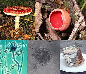
Clockwise from top left:
| |
| Scientific classification | |
| Domain: | Eukaryota |
| Clade: | Obazoa |
| (unranked): | Opisthokonta |
| Clade: | Holomycota |
| Kingdom: | Fungi R.T.Moore (1980)[1][2] |
| Subkingdoms/phyla | |
Грибок Грибы ( пл .: [ 3 ] или грибки [ 4 ] ) является любым членом группы эукариотических организмов, которая включает в себя микроорганизмы, такие как дрожжи и плесени , а также более знакомые грибы . Эти организмы классифицируются как одно из традиционных эукариотических королевств , наряду с животными , платья и либо профиста [ 5 ] или простейшие и хромиста . [ 6 ]
A characteristic that places fungi in a different kingdom from plants, bacteria, and some protists is chitin in their cell walls. Fungi, like animals, are heterotrophs; they acquire their food by absorbing dissolved molecules, typically by secreting digestive enzymes into their environment. Fungi do not photosynthesize. Growth is their means of mobility, except for spores (a few of which are flagellated), which may travel through the air or water. Fungi are the principal decomposers in ecological systems. These and other differences place fungi in a single group of related organisms, named the Eumycota (true fungi or Eumycetes), that share a common ancestor (i.e. they form a monophyletic group), an interpretation that is also strongly supported by molecular phylogenetics. This fungal group is distinct from the structurally similar myxomycetes (slime molds) and oomycetes (water molds). The discipline of biology devoted to the study of fungi is known as mycology (from the Greek μύκης mykes, mushroom). In the past, mycology was regarded as a branch of Ботаника , хотя теперь известно, что грибы генетически более тесно связаны с животными, чем с растениями.
Abundant worldwide, most fungi are inconspicuous because of the small size of their structures, and their cryptic lifestyles in soil or on dead matter. Fungi include symbionts of plants, animals, or other fungi and also parasites. They may become noticeable when fruiting, either as mushrooms or as molds. Fungi perform an essential role in the decomposition of organic matter and have fundamental roles in nutrient cycling and exchange in the environment. They have long been used as a direct source of human food, in the form of mushrooms and truffles; as a leavening agent for bread; and in the fermentation of various food products, such as wine, beer, and soy sauce. Since the 1940s, fungi have been used for the production of antibiotics, and, more recently, various enzymes produced by fungi are used industrially and in detergents. Fungi are also used as biological pesticides to control weeds, plant diseases, and insect pests. Many species produce bioactive compounds called mycotoxins, such as alkaloids and polyketides, that are toxic to animals, including humans. The fruiting structures of a few species contain psychotropic compounds and are consumed recreationally or in traditional spiritual ceremonies. Fungi can break down manufactured materials and buildings, and become significant pathogens of humans and other animals. Losses of crops due to fungal diseases (e.g., rice blast disease) or food spoilage can have a large impact on human food supplies and local economies.
The fungus kingdom encompasses an enormous diversity of taxa with varied ecologies, life cycle strategies, and morphologies ranging from unicellular aquatic chytrids to large mushrooms. However, little is known of the true biodiversity of the fungus kingdom, which has been estimated at 2.2 million to 3.8 million species.[7] Of these, only about 148,000 have been described,[8] with over 8,000 species known to be detrimental to plants and at least 300 that can be pathogenic to humans.[9] Ever since the pioneering 18th and 19th century taxonomical works of Carl Linnaeus, Christiaan Hendrik Persoon, and Elias Magnus Fries, fungi have been classified according to their morphology (e.g., characteristics such as spore color or microscopic features) or physiology. Advances in molecular genetics have opened the way for DNA analysis to be incorporated into taxonomy, which has sometimes challenged the historical groupings based on morphology and other traits. Phylogenetic studies published in the first decade of the 21st century have helped reshape the classification within the fungi kingdom, which is divided into one subkingdom, seven phyla, and ten subphyla.
Etymology
The English word fungus is directly adopted from the Latin fungus (mushroom), used in the writings of Horace and Pliny.[10] This in turn is derived from the Greek word sphongos (σφόγγος 'sponge'), which refers to the macroscopic structures and morphology of mushrooms and molds;[11] the root is also used in other languages, such as the German Schwamm ('sponge') and Schimmel ('mold').[12]
The word mycology is derived from the Greek mykes (μύκης 'mushroom') and logos (λόγος 'discourse').[13] It denotes the scientific study of fungi. The Latin adjectival form of "mycology" (mycologicæ) appeared as early as 1796 in a book on the subject by Christiaan Hendrik Persoon.[14] The word appeared in English as early as 1824 in a book by Robert Kaye Greville.[15] In 1836 the English naturalist Miles Joseph Berkeley's publication The English Flora of Sir James Edward Smith, Vol. 5. also refers to mycology as the study of fungi.[11][16]
A group of all the fungi present in a particular region is known as mycobiota (plural noun, no singular).[17] The term mycota is often used for this purpose, but many authors use it as a synonym of Fungi. The word funga has been proposed as a less ambiguous term morphologically similar to fauna and flora.[18] The Species Survival Commission (SSC) of the International Union for Conservation of Nature (IUCN) in August 2021 asked that the phrase fauna and flora be replaced by fauna, flora, and funga.[19]
Characteristics


Before the introduction of molecular methods for phylogenetic analysis, taxonomists considered fungi to be members of the plant kingdom because of similarities in lifestyle: both fungi and plants are mainly immobile, and have similarities in general morphology and growth habitat. Although inaccurate, the common misconception that fungi are plants persists among the general public due to their historical classification, as well as several similarities.[20][21] Like plants, fungi often grow in soil and, in the case of mushrooms, form conspicuous fruit bodies, which sometimes resemble plants such as mosses. The fungi are now considered a separate kingdom, distinct from both plants and animals, from which they appear to have diverged around one billion years ago (around the start of the Neoproterozoic Era).[22][23] Some morphological, biochemical, and genetic features are shared with other organisms, while others are unique to the fungi, clearly separating them from the other kingdoms:
Shared features:
- With other eukaryotes: Fungal cells contain membrane-bound nuclei with chromosomes that contain DNA with noncoding regions called introns and coding regions called exons. Fungi have membrane-bound cytoplasmic organelles such as mitochondria, sterol-containing membranes, and ribosomes of the 80S type.[24] They have a characteristic range of soluble carbohydrates and storage compounds, including sugar alcohols (e.g., mannitol), disaccharides, (e.g., trehalose), and polysaccharides (e.g., glycogen, which is also found in animals[25]).
- With animals: Fungi lack chloroplasts and are heterotrophic organisms and so require preformed organic compounds as energy sources.[26]
- With plants: Fungi have a cell wall[27] and vacuoles.[28] They reproduce by both sexual and asexual means, and like basal plant groups (such as ferns and mosses) produce spores. Similar to mosses and algae, fungi typically have haploid nuclei.[29]
- With euglenoids and bacteria: Higher fungi, euglenoids, and some bacteria produce the amino acid L-lysine in specific biosynthesis steps, called the α-aminoadipate pathway.[30][31]
- The cells of most fungi grow as tubular, elongated, and thread-like (filamentous) structures called hyphae, which may contain multiple nuclei and extend by growing at their tips. Each tip contains a set of aggregated vesicles—cellular structures consisting of proteins, lipids, and other organic molecules—called the Spitzenkörper.[32] Both fungi and oomycetes grow as filamentous hyphal cells.[33] In contrast, similar-looking organisms, such as filamentous green algae, grow by repeated cell division within a chain of cells.[25] There are also single-celled fungi (yeasts) that do not form hyphae, and some fungi have both hyphal and yeast forms.[34]
- In common with some plant and animal species, more than one hundred fungal species display bioluminescence.[35]
Unique features:
- Some species grow as unicellular yeasts that reproduce by budding or fission. Dimorphic fungi can switch between a yeast phase and a hyphal phase in response to environmental conditions.[34]
- The fungal cell wall is made of a chitin-glucan complex; while glucans are also found in plants and chitin in the exoskeleton of arthropods,[36] fungi are the only organisms that combine these two structural molecules in their cell wall. Unlike those of plants and oomycetes, fungal cell walls do not contain cellulose.[37][38]
Most fungi lack an efficient system for the long-distance transport of water and nutrients, such as the xylem and phloem in many plants. To overcome this limitation, some fungi, such as Armillaria, form rhizomorphs,[39] which resemble and perform functions similar to the roots of plants. As eukaryotes, fungi possess a biosynthetic pathway for producing terpenes that uses mevalonic acid and pyrophosphate as chemical building blocks.[40] Plants and some other organisms have an additional terpene biosynthesis pathway in their chloroplasts, a structure that fungi and animals do not have.[41] Fungi produce several secondary metabolites that are similar or identical in structure to those made by plants.[40] Many of the plant and fungal enzymes that make these compounds differ from each other in sequence and other characteristics, which indicates separate origins and convergent evolution of these enzymes in the fungi and plants.[40][42]
Diversity
Fungi have a worldwide distribution, and grow in a wide range of habitats, including extreme environments such as deserts or areas with high salt concentrations[43] or ionizing radiation,[44] as well as in deep sea sediments.[45] Some can survive the intense UV and cosmic radiation encountered during space travel.[46] Most grow in terrestrial environments, though several species live partly or solely in aquatic habitats, such as the chytrid fungi Batrachochytrium dendrobatidis and B. salamandrivorans, parasites that have been responsible for a worldwide decline in amphibian populations. These organisms spend part of their life cycle as a motile zoospore, enabling them to propel themselves through water and enter their amphibian host.[47] Other examples of aquatic fungi include those living in hydrothermal areas of the ocean.[48]

As of 2020,[update] around 148,000 species of fungi have been described by taxonomists,[8] but the global biodiversity of the fungus kingdom is not fully understood.[50] A 2017 estimate suggests there may be between 2.2 and 3.8 million species.[7] The number of new fungi species discovered yearly has increased from 1,000 to 1,500 per year about 10 years ago, to about 2,000 with a peak of more than 2,500 species in 2016. In the year 2019, 1,882 new species of fungi were described, and it was estimated that more than 90% of fungi remain unknown.[8] The following year, 2,905 new species were described—the highest annual record of new fungus names.[51] In mycology, species have historically been distinguished by a variety of methods and concepts. Classification based on morphological characteristics, such as the size and shape of spores or fruiting structures, has traditionally dominated fungal taxonomy.[52] Species may also be distinguished by their biochemical and physiological characteristics, such as their ability to metabolize certain biochemicals, or their reaction to chemical tests. The biological species concept discriminates species based on their ability to mate. The application of molecular tools, such as DNA sequencing and phylogenetic analysis, to study diversity has greatly enhanced the resolution and added robustness to estimates of genetic diversity within various taxonomic groups.[53]
Mycology
Mycology is the branch of biology concerned with the systematic study of fungi, including their genetic and biochemical properties, their taxonomy, and their use to humans as a source of medicine, food, and psychotropic substances consumed for religious purposes, as well as their dangers, such as poisoning or infection. The field of phytopathology, the study of plant diseases, is closely related because many plant pathogens are fungi.[54]
The use of fungi by humans dates back to prehistory; Ötzi the Iceman, a well-preserved mummy of a 5,300-year-old Neolithic man found frozen in the Austrian Alps, carried two species of polypore mushrooms that may have been used as tinder (Fomes fomentarius), or for medicinal purposes (Piptoporus betulinus).[55] Ancient peoples have used fungi as food sources—often unknowingly—for millennia, in the preparation of leavened bread and fermented juices. Some of the oldest written records contain references to the destruction of crops that were probably caused by pathogenic fungi.[56]
History
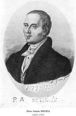
Mycology became a systematic science after the development of the microscope in the 17th century. Although fungal spores were first observed by Giambattista della Porta in 1588, the seminal work in the development of mycology is considered to be the publication of Pier Antonio Micheli's 1729 work Nova plantarum genera.[57] Micheli not only observed spores but also showed that, under the proper conditions, they could be induced into growing into the same species of fungi from which they originated.[58] Extending the use of the binomial system of nomenclature introduced by Carl Linnaeus in his Species plantarum (1753), the Dutch Christiaan Hendrik Persoon (1761–1836) established the first classification of mushrooms with such skill as to be considered a founder of modern mycology. Later, Elias Magnus Fries (1794–1878) further elaborated the classification of fungi, using spore color and microscopic characteristics, methods still used by taxonomists today. Other notable early contributors to mycology in the 17th–19th and early 20th centuries include Miles Joseph Berkeley, August Carl Joseph Corda, Anton de Bary, the brothers Louis René and Charles Tulasne, Arthur H. R. Buller, Curtis G. Lloyd, and Pier Andrea Saccardo. In the 20th and 21st centuries, advances in biochemistry, genetics, molecular biology, biotechnology, DNA sequencing and phylogenetic analysis has provided new insights into fungal relationships and biodiversity, and has challenged traditional morphology-based groupings in fungal taxonomy.[59]
Morphology
Microscopic structures
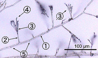
Most fungi grow as hyphae, which are cylindrical, thread-like structures 2–10 μm in diameter and up to several centimeters in length. Hyphae grow at their tips (apices); new hyphae are typically formed by emergence of new tips along existing hyphae by a process called branching, or occasionally growing hyphal tips fork, giving rise to two parallel-growing hyphae.[60] Hyphae also sometimes fuse when they come into contact, a process called hyphal fusion (or anastomosis). These growth processes lead to the development of a mycelium, an interconnected network of hyphae.[34] Hyphae can be either septate or coenocytic. Septate hyphae are divided into compartments separated by cross walls (internal cell walls, called septa, that are formed at right angles to the cell wall giving the hypha its shape), with each compartment containing one or more nuclei; coenocytic hyphae are not compartmentalized.[61] Septa have pores that allow cytoplasm, organelles, and sometimes nuclei to pass through; an example is the dolipore septum in fungi of the phylum Basidiomycota.[62] Coenocytic hyphae are in essence multinucleate supercells.[63]
Many species have developed specialized hyphal structures for nutrient uptake from living hosts; examples include haustoria in plant-parasitic species of most fungal phyla,[64] and arbuscules of several mycorrhizal fungi, which penetrate into the host cells to consume nutrients.[65]
Although fungi are opisthokonts—a grouping of evolutionarily related organisms broadly characterized by a single posterior flagellum—all phyla except for the chytrids have lost their posterior flagella.[66] Fungi are unusual among the eukaryotes in having a cell wall that, in addition to glucans (e.g., β-1,3-glucan) and other typical components, also contains the biopolymer chitin.[38]
Macroscopic structures
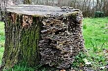
Fungal mycelia can become visible to the naked eye, for example, on various surfaces and substrates, such as damp walls and spoiled food, where they are commonly called molds. Mycelia grown on solid agar media in laboratory petri dishes are usually referred to as colonies. These colonies can exhibit growth shapes and colors (due to spores or pigmentation) that can be used as diagnostic features in the identification of species or groups.[67] Some individual fungal colonies can reach extraordinary dimensions and ages as in the case of a clonal colony of Armillaria solidipes, which extends over an area of more than 900 ha (3.5 square miles), with an estimated age of nearly 9,000 years.[68]
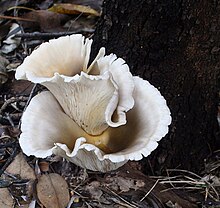
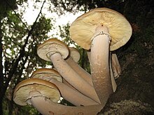
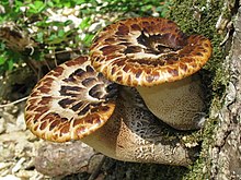
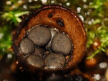
The apothecium—a specialized structure important in sexual reproduction in the ascomycetes—is a cup-shaped fruit body that is often macroscopic and holds the hymenium, a layer of tissue containing the spore-bearing cells.[69] The fruit bodies of the basidiomycetes (basidiocarps) and some ascomycetes can sometimes grow very large, and many are well known as mushrooms.
Growth and physiology
The growth of fungi as hyphae on or in solid substrates or as single cells in aquatic environments is adapted for the efficient extraction of nutrients, because these growth forms have high surface area to volume ratios.[70] Hyphae are specifically adapted for growth on solid surfaces, and to invade substrates and tissues.[71] They can exert large penetrative mechanical forces; for example, many plant pathogens, including Magnaporthe grisea, form a structure called an appressorium that evolved to puncture plant tissues.[72] The pressure generated by the appressorium, directed against the plant epidermis, can exceed 8 megapascals (1,200 psi).[72] The filamentous fungus Paecilomyces lilacinus uses a similar structure to penetrate the eggs of nematodes.[73]
The mechanical pressure exerted by the appressorium is generated from physiological processes that increase intracellular turgor by producing osmolytes such as glycerol.[74] Adaptations such as these are complemented by hydrolytic enzymes secreted into the environment to digest large organic molecules—such as polysaccharides, proteins, and lipids—into smaller molecules that may then be absorbed as nutrients.[75][76][77] The vast majority of filamentous fungi grow in a polar fashion (extending in one direction) by elongation at the tip (apex) of the hypha.[78] Other forms of fungal growth include intercalary extension (longitudinal expansion of hyphal compartments that are below the apex) as in the case of some endophytic fungi,[79] or growth by volume expansion during the development of mushroom stipes and other large organs.[80] Growth of fungi as multicellular structures consisting of somatic and reproductive cells—a feature independently evolved in animals and plants[81]—has several functions, including the development of fruit bodies for dissemination of sexual spores (see above) and biofilms for substrate colonization and intercellular communication.[82]
Fungi are traditionally considered heterotrophs, organisms that rely solely on carbon fixed by other organisms for metabolism. Fungi have evolved a high degree of metabolic versatility that allows them to use a diverse range of organic substrates for growth, including simple compounds such as nitrate, ammonia, acetate, or ethanol.[83][84] In some species the pigment melanin may play a role in extracting energy from ionizing radiation, such as gamma radiation. This form of "radiotrophic" growth has been described for only a few species, the effects on growth rates are small, and the underlying biophysical and biochemical processes are not well known.[44] This process might bear similarity to CO2 fixation via visible light, but instead uses ionizing radiation as a source of energy.[85]
Reproduction
Fungal reproduction is complex, reflecting the differences in lifestyles and genetic makeup within this diverse kingdom of organisms.[86] It is estimated that a third of all fungi reproduce using more than one method of propagation; for example, reproduction may occur in two well-differentiated stages within the life cycle of a species, the teleomorph (sexual reproduction) and the anamorph (asexual reproduction).[87] Environmental conditions trigger genetically determined developmental states that lead to the creation of specialized structures for sexual or asexual reproduction. These structures aid reproduction by efficiently dispersing spores or spore-containing propagules.
Asexual reproduction
Asexual reproduction occurs via vegetative spores (conidia) or through mycelial fragmentation. Mycelial fragmentation occurs when a fungal mycelium separates into pieces, and each component grows into a separate mycelium. Mycelial fragmentation and vegetative spores maintain clonal populations adapted to a specific niche, and allow more rapid dispersal than sexual reproduction.[88] The "Fungi imperfecti" (fungi lacking the perfect or sexual stage) or Deuteromycota comprise all the species that lack an observable sexual cycle.[89] Deuteromycota (alternatively known as Deuteromycetes, conidial fungi, or mitosporic fungi) is not an accepted taxonomic clade and is now taken to mean simply fungi that lack a known sexual stage.[90]
Sexual reproduction
Sexual reproduction with meiosis has been directly observed in all fungal phyla except Glomeromycota[91] (genetic analysis suggests meiosis in Glomeromycota as well). It differs in many aspects from sexual reproduction in animals or plants. Differences also exist between fungal groups and can be used to discriminate species by morphological differences in sexual structures and reproductive strategies.[92][93] Mating experiments between fungal isolates may identify species on the basis of biological species concepts.[93] The major fungal groupings have initially been delineated based on the morphology of their sexual structures and spores; for example, the spore-containing structures, asci and basidia, can be used in the identification of ascomycetes and basidiomycetes, respectively. Fungi employ two mating systems: heterothallic species allow mating only between individuals of the opposite mating type, whereas homothallic species can mate, and sexually reproduce, with any other individual or itself.[94]
Most fungi have both a haploid and a diploid stage in their life cycles. In sexually reproducing fungi, compatible individuals may combine by fusing their hyphae together into an interconnected network; this process, anastomosis, is required for the initiation of the sexual cycle. Many ascomycetes and basidiomycetes go through a dikaryotic stage, in which the nuclei inherited from the two parents do not combine immediately after cell fusion, but remain separate in the hyphal cells (see heterokaryosis).[95]
In ascomycetes, dikaryotic hyphae of the hymenium (the spore-bearing tissue layer) form a characteristic hook (crozier) at the hyphal septum. During cell division, the formation of the hook ensures proper distribution of the newly divided nuclei into the apical and basal hyphal compartments. An ascus (plural asci) is then formed, in which karyogamy (nuclear fusion) occurs. Asci are embedded in an ascocarp, or fruiting body. Karyogamy in the asci is followed immediately by meiosis and the production of ascospores. After dispersal, the ascospores may germinate and form a new haploid mycelium.[96]
Sexual reproduction in basidiomycetes is similar to that of the ascomycetes. Compatible haploid hyphae fuse to produce a dikaryotic mycelium. However, the dikaryotic phase is more extensive in the basidiomycetes, often also present in the vegetatively growing mycelium. A specialized anatomical structure, called a clamp connection, is formed at each hyphal septum. As with the structurally similar hook in the ascomycetes, the clamp connection in the basidiomycetes is required for controlled transfer of nuclei during cell division, to maintain the dikaryotic stage with two genetically different nuclei in each hyphal compartment.[97] A basidiocarp is formed in which club-like structures known as basidia generate haploid basidiospores after karyogamy and meiosis.[98] The most commonly known basidiocarps are mushrooms, but they may also take other forms (see Morphology section).
In fungi formerly classified as Zygomycota, haploid hyphae of two individuals fuse, forming a gametangium, a specialized cell structure that becomes a fertile gamete-producing cell. The gametangium develops into a zygospore, a thick-walled spore formed by the union of gametes. When the zygospore germinates, it undergoes meiosis, generating new haploid hyphae, which may then form asexual sporangiospores. These sporangiospores allow the fungus to rapidly disperse and germinate into new genetically identical haploid fungal mycelia.[99]
Spore dispersal
The spores of most of the researched species of fungi are transported by wind.[100][101] Such species often produce dry or hydrophobic spores that do not absorb water and are readily scattered by raindrops, for example.[100][102][103] In other species, both asexual and sexual spores or sporangiospores are often actively dispersed by forcible ejection from their reproductive structures. This ejection ensures exit of the spores from the reproductive structures as well as traveling through the air over long distances.
Specialized mechanical and physiological mechanisms, as well as spore surface structures (such as hydrophobins), enable efficient spore ejection.[104] For example, the structure of the spore-bearing cells in some ascomycete species is such that the buildup of substances affecting cell volume and fluid balance enables the explosive discharge of spores into the air.[105] The forcible discharge of single spores termed ballistospores involves formation of a small drop of water (Buller's drop), which upon contact with the spore leads to its projectile release with an initial acceleration of more than 10,000 g;[106] the net result is that the spore is ejected 0.01–0.02 cm, sufficient distance for it to fall through the gills or pores into the air below.[107] Other fungi, like the puffballs, rely on alternative mechanisms for spore release, such as external mechanical forces. The hydnoid fungi (tooth fungi) produce spores on pendant, tooth-like or spine-like projections.[108] The bird's nest fungi use the force of falling water drops to liberate the spores from cup-shaped fruiting bodies.[109] Another strategy is seen in the stinkhorns, a group of fungi with lively colors and putrid odor that attract insects to disperse their spores.[110]
Homothallism
In homothallic sexual reproduction, two haploid nuclei derived from the same individual fuse to form a zygote that can then undergo meiosis. Homothallic fungi include species with an Aspergillus-like asexual stage (anamorphs) occurring in numerous different genera,[111] several species of the ascomycete genus Cochliobolus,[112] and the ascomycete Pneumocystis jirovecii.[113] The earliest mode of sexual reproduction among eukaryotes was likely homothallism, that is, self-fertile unisexual reproduction.[114]
Other sexual processes
Besides regular sexual reproduction with meiosis, certain fungi, such as those in the genera Penicillium and Aspergillus, may exchange genetic material via parasexual processes, initiated by anastomosis between hyphae and plasmogamy of fungal cells.[115] The frequency and relative importance of parasexual events is unclear and may be lower than other sexual processes. It is known to play a role in intraspecific hybridization[116] and is likely required for hybridization between species, which has been associated with major events in fungal evolution.[117]
Evolution
In contrast to plants and animals, the early fossil record of the fungi is meager. Factors that likely contribute to the under-representation of fungal species among fossils include the nature of fungal fruiting bodies, which are soft, fleshy, and easily degradable tissues, and the microscopic dimensions of most fungal structures, which therefore are not readily evident. Fungal fossils are difficult to distinguish from those of other microbes, and are most easily identified when they resemble extant fungi.[118] Often recovered from a permineralized plant or animal host, these samples are typically studied by making thin-section preparations that can be examined with light microscopy or transmission electron microscopy.[119] Researchers study compression fossils by dissolving the surrounding matrix with acid and then using light or scanning electron microscopy to examine surface details.[120]

The earliest fossils possessing features typical of fungi date to the Paleoproterozoic era, some 2,400 million years ago (Ma); these multicellular benthic organisms had filamentous structures capable of anastomosis.[121] Other studies (2009) estimate the arrival of fungal organisms at about 760–1060 Ma on the basis of comparisons of the rate of evolution in closely related groups.[122] The oldest fossilizied mycelium to be identified from its molecular composition is between 715 and 810 million years old.[123] For much of the Paleozoic Era (542–251 Ma), the fungi appear to have been aquatic and consisted of organisms similar to the extant chytrids in having flagellum-bearing spores.[124] The evolutionary adaptation from an aquatic to a terrestrial lifestyle necessitated a diversification of ecological strategies for obtaining nutrients, including parasitism, saprobism, and the development of mutualistic relationships such as mycorrhiza and lichenization.[125] Studies suggest that the ancestral ecological state of the Ascomycota was saprobism, and that independent lichenization events have occurred multiple times.[126]
In May 2019, scientists reported the discovery of a fossilized fungus, named Ourasphaira giraldae, in the Canadian Arctic, that may have grown on land a billion years ago, well before plants were living on land.[127][128][129] Pyritized fungus-like microfossils preserved in the basal Ediacaran Doushantuo Formation (~635 Ma) have been reported in South China.[130] Earlier, it had been presumed that the fungi colonized the land during the Cambrian (542–488.3 Ma), also long before land plants.[131] Fossilized hyphae and spores recovered from the Ordovician of Wisconsin (460 Ma) resemble modern-day Glomerales, and existed at a time when the land flora likely consisted of only non-vascular bryophyte-like plants.[132] Prototaxites, which was probably a fungus or lichen, would have been the tallest organism of the late Silurian and early Devonian. Fungal fossils do not become common and uncontroversial until the early Devonian (416–359.2 Ma), when they occur abundantly in the Rhynie chert, mostly as Zygomycota and Chytridiomycota.[131][133][134] At about this same time, approximately 400 Ma, the Ascomycota and Basidiomycota diverged,[135] and all modern classes of fungi were present by the Late Carboniferous (Pennsylvanian, 318.1–299 Ma).[136]
Lichens formed a component of the early terrestrial ecosystems, and the estimated age of the oldest terrestrial lichen fossil is 415 Ma;[137] this date roughly corresponds to the age of the oldest known sporocarp fossil, a Paleopyrenomycites species found in the Rhynie Chert.[138] The oldest fossil with microscopic features resembling modern-day basidiomycetes is Palaeoancistrus, found permineralized with a fern from the Pennsylvanian.[139] Rare in the fossil record are the Homobasidiomycetes (a taxon roughly equivalent to the mushroom-producing species of the Agaricomycetes). Two amber-preserved specimens provide evidence that the earliest known mushroom-forming fungi (the extinct species Archaeomarasmius leggetti) appeared during the late Cretaceous, 90 Ma.[140][141]
Some time after the Permian–Triassic extinction event (251.4 Ma), a fungal spike (originally thought to be an extraordinary abundance of fungal spores in sediments) formed, suggesting that fungi were the dominant life form at this time, representing nearly 100% of the available fossil record for this period.[142] However, the relative proportion of fungal spores relative to spores formed by algal species is difficult to assess,[143] the spike did not appear worldwide,[144][145] and in many places it did not fall on the Permian–Triassic boundary.[146]
Sixty-five million years ago, immediately after the Cretaceous–Paleogene extinction event that famously killed off most dinosaurs, there was a dramatic increase in evidence of fungi; apparently the death of most plant and animal species led to a huge fungal bloom like "a massive compost heap".[147]
Taxonomy
Although commonly included in botany curricula and textbooks, fungi are more closely related to animals than to plants and are placed with the animals in the monophyletic group of opisthokonts.[148] Analyses using molecular phylogenetics support a monophyletic origin of fungi.[53][149] The taxonomy of fungi is in a state of constant flux, especially due to research based on DNA comparisons. These current phylogenetic analyses often overturn classifications based on older and sometimes less discriminative methods based on morphological features and biological species concepts obtained from experimental matings.[150]
There is no unique generally accepted system at the higher taxonomic levels and there are frequent name changes at every level, from species upwards. Efforts among researchers are now underway to establish and encourage usage of a unified and more consistent nomenclature.[53][151] Until relatively recent (2012) changes to the International Code of Nomenclature for algae, fungi and plants, fungal species could also have multiple scientific names depending on their life cycle and mode (sexual or asexual) of reproduction.[152] Web sites such as Index Fungorum and MycoBank are officially recognized nomenclatural repositories and list current names of fungal species (with cross-references to older synonyms).[153]
The 2007 classification of Kingdom Fungi is the result of a large-scale collaborative research effort involving dozens of mycologists and other scientists working on fungal taxonomy.[53] It recognizes seven phyla, two of which—the Ascomycota and the Basidiomycota—are contained within a branch representing subkingdom Dikarya, the most species rich and familiar group, including all the mushrooms, most food-spoilage molds, most plant pathogenic fungi, and the beer, wine, and bread yeasts. The accompanying cladogram depicts the major fungal taxa and their relationship to opisthokont and unikont organisms, based on the work of Philippe Silar,[154] "The Mycota: A Comprehensive Treatise on Fungi as Experimental Systems for Basic and Applied Research"[155] and Tedersoo et al. 2018.[156] The lengths of the branches are not proportional to evolutionary distances.
| Zoosporia |
| ||||||||||||||||||||||||||||||
Taxonomic groups
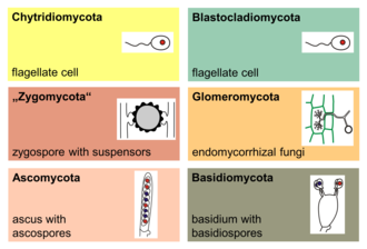
The major phyla (sometimes called divisions) of fungi have been classified mainly on the basis of characteristics of their sexual reproductive structures. As of 2019[update], nine major lineages have been identified: Opisthosporidia, Chytridiomycota, Neocallimastigomycota, Blastocladiomycota, Zoopagomycotina, Mucoromycota, Glomeromycota, Ascomycota and Basidiomycota.[157]
Phylogenetic analysis has demonstrated that the Microsporidia, unicellular parasites of animals and protists, are fairly recent and highly derived endobiotic fungi (living within the tissue of another species).[124] Previously considered to be "primitive" protozoa, they are now thought to be either a basal branch of the Fungi, or a sister group–each other's closest evolutionary relative.[158]
The Chytridiomycota are commonly known as chytrids. These fungi are distributed worldwide. Chytrids and their close relatives Neocallimastigomycota and Blastocladiomycota (below) are the only fungi with active motility, producing zoospores that are capable of active movement through aqueous phases with a single flagellum, leading early taxonomists to classify them as protists. Molecular phylogenies, inferred from rRNA sequences in ribosomes, suggest that the Chytrids are a basal group divergent from the other fungal phyla, consisting of four major clades with suggestive evidence for paraphyly or possibly polyphyly.[159]
The Blastocladiomycota were previously considered a taxonomic clade within the Chytridiomycota. Molecular data and ultrastructural characteristics, however, place the Blastocladiomycota as a sister clade to the Zygomycota, Glomeromycota, and Dikarya (Ascomycota and Basidiomycota). The blastocladiomycetes are saprotrophs, feeding on decomposing organic matter, and they are parasites of all eukaryotic groups. Unlike their close relatives, the chytrids, most of which exhibit zygotic meiosis, the blastocladiomycetes undergo sporic meiosis.[124]
The Neocallimastigomycota were earlier placed in the phylum Chytridiomycota. Members of this small phylum are anaerobic organisms, living in the digestive system of larger herbivorous mammals and in other terrestrial and aquatic environments enriched in cellulose (e.g., domestic waste landfill sites).[160] They lack mitochondria but contain hydrogenosomes of mitochondrial origin. As in the related chrytrids, neocallimastigomycetes form zoospores that are posteriorly uniflagellate or polyflagellate.[53]


Members of the Glomeromycota form arbuscular mycorrhizae, a form of mutualist symbiosis wherein fungal hyphae invade plant root cells and both species benefit from the resulting increased supply of nutrients. All known Glomeromycota species reproduce asexually.[91] The symbiotic association between the Glomeromycota and plants is ancient, with evidence dating to 400 million years ago.[161] Formerly part of the Zygomycota (commonly known as 'sugar' and 'pin' molds), the Glomeromycota were elevated to phylum status in 2001 and now replace the older phylum Zygomycota.[162] Fungi that were placed in the Zygomycota are now being reassigned to the Glomeromycota, or the subphyla incertae sedis Mucoromycotina, Kickxellomycotina, the Zoopagomycotina and the Entomophthoromycotina.[53] Some well-known examples of fungi formerly in the Zygomycota include black bread mold (Rhizopus stolonifer), and Pilobolus species, capable of ejecting spores several meters through the air.[163] Medically relevant genera include Mucor, Rhizomucor, and Rhizopus.[164]
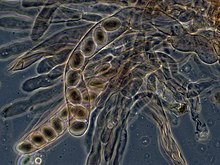
The Ascomycota, commonly known as sac fungi or ascomycetes, constitute the largest taxonomic group within the Eumycota.[52] These fungi form meiotic spores called ascospores, which are enclosed in a special sac-like structure called an ascus. This phylum includes morels, a few mushrooms and truffles, unicellular yeasts (e.g., of the genera Saccharomyces, Kluyveromyces, Pichia, and Candida), and many filamentous fungi living as saprotrophs, parasites, and mutualistic symbionts (e.g. lichens). Prominent and important genera of filamentous ascomycetes include Aspergillus, Penicillium, Fusarium, and Claviceps. Many ascomycete species have only been observed undergoing asexual reproduction (called anamorphic species), but analysis of molecular data has often been able to identify their closest teleomorphs in the Ascomycota.[165] Because the products of meiosis are retained within the sac-like ascus, ascomycetes have been used for elucidating principles of genetics and heredity (e.g., Neurospora crassa).[166]
Members of the Basidiomycota, commonly known as the club fungi or basidiomycetes, produce meiospores called basidiospores on club-like stalks called basidia. Most common mushrooms belong to this group, as well as rust and smut fungi, which are major pathogens of grains. Other important basidiomycetes include the maize pathogen Ustilago maydis,[167] human commensal species of the genus Malassezia,[168] and the opportunistic human pathogen, Cryptococcus neoformans.[169]
Fungus-like organisms
Because of similarities in morphology and lifestyle, the slime molds (mycetozoans, plasmodiophorids, acrasids, Fonticula and labyrinthulids, now in Amoebozoa, Rhizaria, Excavata, Cristidiscoidea and Stramenopiles, respectively), water molds (oomycetes) and hyphochytrids (both Stramenopiles) were formerly classified in the kingdom Fungi, in groups like Mastigomycotina, Gymnomycota and Phycomycetes. The slime molds were studied also as protozoans, leading to an ambiregnal, duplicated taxonomy.[170]
Unlike true fungi, the cell walls of oomycetes contain cellulose and lack chitin. Hyphochytrids have both chitin and cellulose. Slime molds lack a cell wall during the assimilative phase (except labyrinthulids, which have a wall of scales), and take in nutrients by ingestion (phagocytosis, except labyrinthulids) rather than absorption (osmotrophy, as fungi, labyrinthulids, oomycetes and hyphochytrids). Neither water molds nor slime molds are closely related to the true fungi, and, therefore, taxonomists no longer group them in the kingdom Fungi. Nonetheless, studies of the oomycetes and myxomycetes are still often included in mycology textbooks and primary research literature.[171]
The Eccrinales and Amoebidiales are opisthokont protists, previously thought to be zygomycete fungi. Other groups now in Opisthokonta (e.g., Corallochytrium, Ichthyosporea) were also at given time classified as fungi. The genus Blastocystis, now in Stramenopiles, was originally classified as a yeast. Ellobiopsis, now in Alveolata, was considered a chytrid. The bacteria were also included in fungi in some classifications, as the group Schizomycetes.
The Rozellida clade, including the "ex-chytrid" Rozella, is a genetically disparate group known mostly from environmental DNA sequences that is a sister group to fungi.[157] Members of the group that have been isolated lack the chitinous cell wall that is characteristic of fungi. Alternatively, Rozella can be classified as a basal fungal group.[149]
The nucleariids may be the next sister group to the eumycete clade, and as such could be included in an expanded fungal kingdom.[148] Many Actinomycetales (Actinomycetota), a group with many filamentous bacteria, were also long believed to be fungi.[172][173]
Ecology

Although often inconspicuous, fungi occur in every environment on Earth and play very important roles in most ecosystems. Along with bacteria, fungi are the major decomposers in most terrestrial (and some aquatic) ecosystems, and therefore play a critical role in biogeochemical cycles[174] and in many food webs. As decomposers, they play an essential role in nutrient cycling, especially as saprotrophs and symbionts, degrading organic matter to inorganic molecules, which can then re-enter anabolic metabolic pathways in plants or other organisms.[175][176]
Symbiosis
Many fungi have important symbiotic relationships with organisms from most if not all kingdoms.[177][178][179] These interactions can be mutualistic or antagonistic in nature, or in the case of commensal fungi are of no apparent benefit or detriment to the host.[180][181][182]
With plants
Mycorrhizal symbiosis between plants and fungi is one of the most well-known plant–fungus associations and is of significant importance for plant growth and persistence in many ecosystems; over 90% of all plant species engage in mycorrhizal relationships with fungi and are dependent upon this relationship for survival.[183]
The mycorrhizal symbiosis is ancient, dating back to at least 400 million years.[161] It often increases the plant's uptake of inorganic compounds, such as nitrate and phosphate from soils having low concentrations of these key plant nutrients.[175][184] The fungal partners may also mediate plant-to-plant transfer of carbohydrates and other nutrients.[185] Such mycorrhizal communities are called "common mycorrhizal networks".[186][187] A special case of mycorrhiza is myco-heterotrophy, whereby the plant parasitizes the fungus, obtaining all of its nutrients from its fungal symbiont.[188] Some fungal species inhabit the tissues inside roots, stems, and leaves, in which case they are called endophytes.[189] Similar to mycorrhiza, endophytic colonization by fungi may benefit both symbionts; for example, endophytes of grasses impart to their host increased resistance to herbivores and other environmental stresses and receive food and shelter from the plant in return.[190]
With algae and cyanobacteria
Lichens are a symbiotic relationship between fungi and photosynthetic algae or cyanobacteria. The photosynthetic partner in the relationship is referred to in lichen terminology as a "photobiont". The fungal part of the relationship is composed mostly of various species of ascomycetes and a few basidiomycetes.[191] Lichens occur in every ecosystem on all continents, play a key role in soil formation and the initiation of biological succession,[192] and are prominent in some extreme environments, including polar, alpine, and semiarid desert regions.[193] They are able to grow on inhospitable surfaces, including bare soil, rocks, tree bark, wood, shells, barnacles and leaves.[194] As in mycorrhizas, the photobiont provides sugars and other carbohydrates via photosynthesis to the fungus, while the fungus provides minerals and water to the photobiont. The functions of both symbiotic organisms are so closely intertwined that they function almost as a single organism; in most cases the resulting organism differs greatly from the individual components.[195] Lichenization is a common mode of nutrition for fungi; around 27% of known fungi—more than 19,400 species—are lichenized.[196] Characteristics common to most lichens include obtaining organic carbon by photosynthesis, slow growth, small size, long life, long-lasting (seasonal) vegetative reproductive structures, mineral nutrition obtained largely from airborne sources, and greater tolerance of desiccation than most other photosynthetic organisms in the same habitat.[197]
With insects
Many insects also engage in mutualistic relationships with fungi. Several groups of ants cultivate fungi in the order Chaetothyriales for several purposes: as a food source, as a structural component of their nests, and as a part of an ant/plant symbiosis in the domatia (tiny chambers in plants that house arthropods).[198] Ambrosia beetles cultivate various species of fungi in the bark of trees that they infest.[199] Likewise, females of several wood wasp species (genus Sirex) inject their eggs together with spores of the wood-rotting fungus Amylostereum areolatum into the sapwood of pine trees; the growth of the fungus provides ideal nutritional conditions for the development of the wasp larvae.[200] At least one species of stingless bee has a relationship with a fungus in the genus Monascus, where the larvae consume and depend on fungus transferred from old to new nests.[201] Termites on the African savannah are also known to cultivate fungi,[177] and yeasts of the genera Candida and Lachancea inhabit the gut of a wide range of insects, including neuropterans, beetles, and cockroaches; it is not known whether these fungi benefit their hosts.[202] Fungi growing in dead wood are essential for xylophagous insects (e.g. woodboring beetles).[203][204][205] They deliver nutrients needed by xylophages to nutritionally scarce dead wood.[206][204][205] Thanks to this nutritional enrichment the larvae of the woodboring insect is able to grow and develop to adulthood.[203] The larvae of many families of fungicolous flies, particularly those within the superfamily Sciaroidea such as the Mycetophilidae and some Keroplatidae feed on fungal fruiting bodies and sterile mycorrhizae.[207]
As pathogens and parasites

Many fungi are parasites on plants, animals (including humans), and other fungi. Serious pathogens of many cultivated plants causing extensive damage and losses to agriculture and forestry include the rice blast fungus Magnaporthe oryzae,[208] tree pathogens such as Ophiostoma ulmi and Ophiostoma novo-ulmi causing Dutch elm disease,[209] Cryphonectria parasitica responsible for chestnut blight,[210] and Phymatotrichopsis omnivora causing Texas Root Rot, and plant pathogens in the genera Fusarium, Ustilago, Alternaria, and Cochliobolus.[181] Some carnivorous fungi, like Paecilomyces lilacinus, are predators of nematodes, which they capture using an array of specialized structures such as constricting rings or adhesive nets.[211] Many fungi that are plant pathogens, such as Magnaporthe oryzae, can switch from being biotrophic (parasitic on living plants) to being necrotrophic (feeding on the dead tissues of plants they have killed).[212] This same principle is applied to fungi-feeding parasites, including Asterotremella albida, which feeds on the fruit bodies of other fungi both while they are living and after they are dead.[213]
Some fungi can cause serious diseases in humans, several of which may be fatal if untreated. These include aspergillosis, candidiasis, coccidioidomycosis, cryptococcosis, histoplasmosis, mycetomas, and paracoccidioidomycosis. Furthermore, a person with immunodeficiency is more susceptible to disease by genera such as Aspergillus, Candida, Cryptoccocus,[182][214][215] Histoplasma,[216] and Pneumocystis.[217] Other fungi can attack eyes, nails, hair, and especially skin, the so-called dermatophytic and keratinophilic fungi, and cause local infections such as ringworm and athlete's foot.[218] Fungal spores are also a cause of allergies, and fungi from different taxonomic groups can evoke allergic reactions.[219]
As targets of mycoparasites
Organisms that parasitize fungi are known as mycoparasitic organisms. About 300 species of fungi and fungus-like organisms, belonging to 13 classes and 113 genera, are used as biocontrol agents against plant fungal diseases.[220] Fungi can also act as mycoparasites or antagonists of other fungi, such as Hypomyces chrysospermus, which grows on bolete mushrooms. Fungi can also become the target of infection by mycoviruses.[221][222]
Communication
There appears to be electrical communication between fungi in word-like components according to spiking characteristics.[223]
Possible impact on climate
According to a study published in the academic journal Current Biology, fungi can soak from the atmosphere around 36% of global fossil fuel greenhouse gas emissions.[224][225]
Mycotoxins
![(6aR,9R)-N-((2R,5S,10aS,10bS)-5-benzyl-10b-hydroxy-2-methyl-3,6-dioxooctahydro-2H-oxazolo[3,2-a] pyrrolo[2,1-c]pyrazin-2-yl)-7-methyl-4,6,6a,7,8,9-hexahydroindolo[4,3-fg] quinoline-9-carboxamide](http://upload.wikimedia.org/wikipedia/commons/thumb/9/94/Ergotamine3.png/220px-Ergotamine3.png)
Many fungi produce biologically active compounds, several of which are toxic to animals or plants and are therefore called mycotoxins. Of particular relevance to humans are mycotoxins produced by molds causing food spoilage, and poisonous mushrooms (see above). Particularly infamous are the lethal amatoxins in some Amanita mushrooms, and ergot alkaloids, which have a long history of causing serious epidemics of ergotism (St Anthony's Fire) in people consuming rye or related cereals contaminated with sclerotia of the ergot fungus, Claviceps purpurea.[226] Other notable mycotoxins include the aflatoxins, which are insidious liver toxins and highly carcinogenic metabolites produced by certain Aspergillus species often growing in or on grains and nuts consumed by humans, ochratoxins, patulin, and trichothecenes (e.g., T-2 mycotoxin) and fumonisins, which have significant impact on human food supplies or animal livestock.[227]
Mycotoxins are secondary metabolites (or natural products), and research has established the existence of biochemical pathways solely for the purpose of producing mycotoxins and other natural products in fungi.[40] Mycotoxins may provide fitness benefits in terms of physiological adaptation, competition with other microbes and fungi, and protection from consumption (fungivory).[228][229] Many fungal secondary metabolites (or derivatives) are used medically, as described under Human use below.
Pathogenic mechanisms
Ustilago maydis is a pathogenic plant fungus that causes smut disease in maize and teosinte. Plants have evolved efficient defense systems against pathogenic microbes such as U. maydis. A rapid defense reaction after pathogen attack is the oxidative burst where the plant produces reactive oxygen species at the site of the attempted invasion. U. maydis can respond to the oxidative burst with an oxidative stress response, regulated by the gene YAP1. The response protects U. maydis from the host defense, and is necessary for the pathogen's virulence.[230] Furthermore, U. maydis has a well-established recombinational DNA repair system which acts during mitosis and meiosis.[231] The system may assist the pathogen in surviving DNA damage arising from the host plant's oxidative defensive response to infection.[232]
Cryptococcus neoformans is an encapsulated yeast that can live in both plants and animals. C. neoformans usually infects the lungs, where it is phagocytosed by alveolar macrophages.[233] Some C. neoformans can survive inside macrophages, which appears to be the basis for latency, disseminated disease, and resistance to antifungal agents. One mechanism by which C. neoformans survives the hostile macrophage environment is by up-regulating the expression of genes involved in the oxidative stress response.[233] Another mechanism involves meiosis. The majority of C. neoformans are mating "type a". Filaments of mating "type a" ordinarily have haploid nuclei, but they can become diploid (perhaps by endoduplication or by stimulated nuclear fusion) to form blastospores. The diploid nuclei of blastospores can undergo meiosis, including recombination, to form haploid basidiospores that can be dispersed.[234] This process is referred to as monokaryotic fruiting. This process requires a gene called DMC1, which is a conserved homologue of genes recA in bacteria and RAD51 in eukaryotes, that mediates homologous chromosome pairing during meiosis and repair of DNA double-strand breaks. Thus, C. neoformans can undergo a meiosis, monokaryotic fruiting, that promotes recombinational repair in the oxidative, DNA damaging environment of the host macrophage, and the repair capability may contribute to its virulence.[232][234]
Human use

The human use of fungi for food preparation or preservation and other purposes is extensive and has a long history. Mushroom farming and mushroom gathering are large industries in many countries. The study of the historical uses and sociological impact of fungi is known as ethnomycology. Because of the capacity of this group to produce an enormous range of natural products with antimicrobial or other biological activities, many species have long been used or are being developed for industrial production of antibiotics, vitamins, and anti-cancer and cholesterol-lowering drugs. Methods have been developed for genetic engineering of fungi,[235] enabling metabolic engineering of fungal species. For example, genetic modification of yeast species[236]—which are easy to grow at fast rates in large fermentation vessels—has opened up ways of pharmaceutical production that are potentially more efficient than production by the original source organisms.[237] Fungi-based industries are sometimes considered to be a major part of a growing bioeconomy, with applications under research and development including use for textiles, meat substitution and general fungal biotechnology.[238][239][240][241][242]
Therapeutic uses

Modern chemotherapeutics
Many species produce metabolites that are major sources of pharmacologically active drugs.
Antibiotics
Particularly important are the antibiotics, including the penicillins, a structurally related group of β-lactam antibiotics that are synthesized from small peptides. Although naturally occurring penicillins such as penicillin G (produced by Penicillium chrysogenum) have a relatively narrow spectrum of biological activity, a wide range of other penicillins can be produced by chemical modification of the natural penicillins. Modern penicillins are semisynthetic compounds, obtained initially from fermentation cultures, but then structurally altered for specific desirable properties.[244] Other antibiotics produced by fungi include: ciclosporin, commonly used as an immunosuppressant during transplant surgery; and fusidic acid, used to help control infection from methicillin-resistant Staphylococcus aureus bacteria.[245] Widespread use of antibiotics for the treatment of bacterial diseases, such as tuberculosis, syphilis, leprosy, and others began in the early 20th century and continues to date. In nature, antibiotics of fungal or bacterial origin appear to play a dual role: at high concentrations they act as chemical defense against competition with other microorganisms in species-rich environments, such as the rhizosphere, and at low concentrations as quorum-sensing molecules for intra- or interspecies signaling.[246]
Other
Other drugs produced by fungi include griseofulvin isolated from Penicillium griseofulvum, used to treat fungal infections,[247] and statins (HMG-CoA reductase inhibitors), used to inhibit cholesterol synthesis. Examples of statins found in fungi include mevastatin from Penicillium citrinum and lovastatin from Aspergillus terreus and the oyster mushroom.[248] Psilocybin from fungi is investigated for therapeutic use and appears to cause global increases in brain network integration.[249] Fungi produce compounds that inhibit viruses[250][251] and cancer cells.[252] Specific metabolites, such as polysaccharide-K, ergotamine, and β-lactam antibiotics, are routinely used in clinical medicine. The shiitake mushroom is a source of lentinan, a clinical drug approved for use in cancer treatments in several countries, including Japan.[253][254] In Europe and Japan, polysaccharide-K (brand name Krestin), a chemical derived from Trametes versicolor, is an approved adjuvant for cancer therapy.[255]
Traditional medicine


Certain mushrooms are used as supposed therapeutics in folk medicine practices, such as traditional Chinese medicine. Mushrooms with a history of such use include Agaricus subrufescens,[252][256] Ganoderma lucidum,[257] and Ophiocordyceps sinensis.[258]
Cultured foods
Baker's yeast or Saccharomyces cerevisiae, a unicellular fungus, is used to make bread and other wheat-based products, such as pizza dough and dumplings.[259] Yeast species of the genus Saccharomyces are also used to produce alcoholic beverages through fermentation.[260] Shoyu koji mold (Aspergillus oryzae) is an essential ingredient in brewing Shoyu (soy sauce) and sake, and the preparation of miso,[261] while Rhizopus species are used for making tempeh.[262] Several of these fungi are domesticated species that were bred or selected according to their capacity to ferment food without producing harmful mycotoxins (see below), which are produced by very closely related Aspergilli.[263] Quorn, a meat substitute, is made from Fusarium venenatum.[264]
In food

Edible mushrooms include commercially raised and wild-harvested fungi. Agaricus bisporus, sold as button mushrooms when small or Portobello mushrooms when larger, is the most widely cultivated species in the West, used in salads, soups, and many other dishes. Many Asian fungi are commercially grown and have increased in popularity in the West. They are often available fresh in grocery stores and markets, including straw mushrooms (Volvariella volvacea), oyster mushrooms (Pleurotus ostreatus), shiitakes (Lentinula edodes), and enokitake (Flammulina spp.).[265]

Many other mushroom species are harvested from the wild for personal consumption or commercial sale. Milk mushrooms, morels, chanterelles, truffles, black trumpets, and porcini mushrooms (Boletus edulis) (also known as king boletes) demand a high price on the market. They are often used in gourmet dishes.[266]
Certain types of cheeses require inoculation of milk curds with fungal species that impart a unique flavor and texture to the cheese. Examples include the blue color in cheeses such as Stilton or Roquefort, which are made by inoculation with Penicillium roqueforti.[267] Molds used in cheese production are non-toxic and are thus safe for human consumption; however, mycotoxins (e.g., aflatoxins, roquefortine C, patulin, or others) may accumulate because of growth of other fungi during cheese ripening or storage.[268]
Poisonous fungi

Many mushroom species are poisonous to humans and cause a range of reactions including slight digestive problems, allergic reactions, hallucinations, severe organ failure, and death. Genera with mushrooms containing deadly toxins include Conocybe, Galerina, Lepiota and the most infamous, Amanita.[269] The latter genus includes the destroying angel (A. virosa) and the death cap (A. phalloides), the most common cause of deadly mushroom poisoning.[270] The false morel (Gyromitra esculenta) is occasionally considered a delicacy when cooked, yet can be highly toxic when eaten raw.[271] Tricholoma equestre was considered edible until it was implicated in serious poisonings causing rhabdomyolysis.[272] Fly agaric mushrooms (Amanita muscaria) also cause occasional non-fatal poisonings, mostly as a result of ingestion for its hallucinogenic properties. Historically, fly agaric was used by different peoples in Europe and Asia and its present usage for religious or shamanic purposes is reported from some ethnic groups such as the Koryak people of northeastern Siberia.[273]
As it is difficult to accurately identify a safe mushroom without proper training and knowledge, it is often advised to assume that a wild mushroom is poisonous and not to consume it.[274][275]
Pest control
In agriculture, fungi may be useful if they actively compete for nutrients and space with pathogenic microorganisms such as bacteria or other fungi via the competitive exclusion principle,[276] or if they are parasites of these pathogens. For example, certain species eliminate or suppress the growth of harmful plant pathogens, such as insects, mites, weeds, nematodes, and other fungi that cause diseases of important crop plants.[277] This has generated strong interest in practical applications that use these fungi in the biological control of these agricultural pests. Entomopathogenic fungi can be used as biopesticides, as they actively kill insects.[278] Examples that have been used as biological insecticides are Beauveria bassiana, Metarhizium spp., Hirsutella spp., Paecilomyces (Isaria) spp., and Lecanicillium lecanii.[279][280] Endophytic fungi of grasses of the genus Epichloë, such as E. coenophiala, produce alkaloids that are toxic to a range of invertebrate and vertebrate herbivores. These alkaloids protect grass plants from herbivory, but several endophyte alkaloids can poison grazing animals, such as cattle and sheep.[281] Infecting cultivars of pasture or forage grasses with Epichloë endophytes is one approach being used in grass breeding programs; the fungal strains are selected for producing only alkaloids that increase resistance to herbivores such as insects, while being non-toxic to livestock.[282][283]
Bioremediation
Certain fungi, in particular white-rot fungi, can degrade insecticides, herbicides, pentachlorophenol, creosote, coal tars, and heavy fuels and turn them into carbon dioxide, water, and basic elements.[284] Fungi have been shown to biomineralize uranium oxides, suggesting they may have application in the bioremediation of radioactively polluted sites.[285][286][287]
Model organisms
Several pivotal discoveries in biology were made by researchers using fungi as model organisms, that is, fungi that grow and sexually reproduce rapidly in the laboratory. For example, the one gene-one enzyme hypothesis was formulated by scientists using the bread mold Neurospora crassa to test their biochemical theories.[288] Other important model fungi are Aspergillus nidulans and the yeasts Saccharomyces cerevisiae and Schizosaccharomyces pombe, each of which with a long history of use to investigate issues in eukaryotic cell biology and genetics, such as cell cycle regulation, chromatin structure, and gene regulation. Other fungal models have emerged that address specific biological questions relevant to medicine, plant pathology, and industrial uses; examples include Candida albicans, a dimorphic, opportunistic human pathogen,[289] Magnaporthe grisea, a plant pathogen,[290] and Pichia pastoris, a yeast widely used for eukaryotic protein production.[291]
Others
Fungi are used extensively to produce industrial chemicals like citric, gluconic, lactic, and malic acids,[292] and industrial enzymes, such as lipases used in biological detergents,[293] cellulases used in making cellulosic ethanol[294] and stonewashed jeans,[295] and amylases,[296] invertases, proteases and xylanases.[297]
See also
References
Citations
- ^ Moore RT (1980). "Taxonomic proposals for the classification of marine yeasts and other yeast-like fungi including the smuts". Botanica Marina. 23 (6): 361–373. doi:10.1515/bot-1980-230605.
- ^ "Record Details: Fungi R.T. Moore, Bot. Mar. 23(6): 371 (1980)". Index Fungorum. Retrieved 18 June 2024.
- ^ /ˈfʌndʒaɪ/ , /ˈfʌŋɡaɪ/ , /ˈfʌŋɡi/ or /ˈfʌndʒi/ . The first two pronunciations are favored more in the US and the others in the UK, however all pronunciations can be heard in any English-speaking country.
- ^ "Fungus". Oxford Dictionaries. Archived from the original on 28 July 2012. Retrieved 26 February 2011.
- ^ Whittaker R (January 1969). "New concepts of kingdoms or organisms. Evolutionary relations are better represented by new classifications than by the traditional two kingdoms". Science. 163 (3863): 150–60. Bibcode:1969Sci...163..150W. CiteSeerX 10.1.1.403.5430. doi:10.1126/science.163.3863.150. PMID 5762760.
- ^ Cavalier-Smith T (1998). "A revised six-kingdom system of life". Biological Reviews. 73 (3): 203–66. doi:10.1111/j.1469-185X.1998.tb00030.x. PMID 9809012. S2CID 6557779.
- ^ Jump up to: a b Hawksworth DL, Lücking R (July 2017). "Fungal Diversity Revisited: 2.2 to 3.8 Million Species". Microbiology Spectrum. 5 (4): 79–95. doi:10.1128/microbiolspec.FUNK-0052-2016. ISBN 978-1-55581-957-6. PMID 28752818.
- ^ Jump up to: a b c Cheek M, Nic Lughadha E, Kirk P, Lindon H, Carretero J, Looney B, et al. (2020). "New scientific discoveries: Plants and fungi". Plants, People, Planet. 2 (5): 371–388. doi:10.1002/ppp3.10148. hdl:1854/LU-8705210.
- ^ "Stop neglecting fungi". Nature Microbiology. 2 (8): 17120. 25 July 2017. doi:10.1038/nmicrobiol.2017.120. PMID 28741610.
- ^ Simpson DP (1979). Cassell's Latin Dictionary (5 ed.). London, UK: Cassell Ltd. p. 883. ISBN 978-0-304-52257-6.
- ^ Jump up to: a b Ainsworth 1976, p. 2.
- ^ Mitzka W, ed. (1960). Etymologisches Wörterbuch der deutschen Sprache [Etymological dictionary of the German language] (in German). Berlin: Walter de Gruyter.
- ^ Alexopoulos, Mims & Blackwell 1996, p. 1.
- ^ Persoon CH (1796). Observationes Mycologicae: Part 1 (in Latin). Leipzig, (Germany): Peter Philipp Wolf. Archived from the original on 19 December 2013. Retrieved 30 March 2019.
- ^ Greville RK (1824). Scottish Cryptogamie Flora: Or Coloured Figures and Descriptions of Cryptogamic Plants, Belonging Chiefly to the Order Fungi. Vol. 2. Edinburgh, Scotland: Maclachland and Stewart. p. 65. From p. 65: "This little plant will probably not prove rare in Great Britain, when mycology shall be more studied."
- ^ Smith JE (1836). Hooker WJ, Berkeley MJ (eds.). The English Flora of Sir James Edward Smith. Vol. 5, part II: "Class XXIV. Cryptogamia". London, England: Longman, Rees, Orme, Brown, Green & Longman. p. 7. From p. 7: "This has arisen, I conceive, partly from the practical difficulty of preserving specimens for the herbarium, partly from the absence of any general work, adapted to the immense advances which have of late years been made in the study of Mycology."
- ^ "LIAS Glossary". Archived from the original on 11 December 2013. Retrieved 14 August 2013.
- ^ Kuhar F, Furci G, Drechsler-Santos ER, Pfister DH (2018). "Delimitation of Funga as a valid term for the diversity of fungal communities: the Fauna, Flora & Funga proposal (FF&F)". IMA Fungus. 9 (2): A71–A74. doi:10.1007/BF03449441. hdl:11336/88035.
- ^ "IUCN SSC acceptance of Fauna Flora Funga" (PDF). Fungal Conservation Committee, IUCN SSC. 2021. Archived from the original (PDF) on 11 November 2021. Retrieved 11 November 2021.
The IUCN Species Survival Commission calls for the due recognition of fungi as major components of biodiversity in legislation and policy. It fully endorses the Fauna Flora Funga Initiative and asks that the phrases animals and plants and fauna and flora be replaced with animals, fungi, and plants and fauna, flora, and funga.
- ^ "Fifth-Grade Elementary School Students' Conceptions and Misconceptions about the Fungus Kingdom". Retrieved 5 October 2022.
- ^ "Common Student Ideas about Plants and Animals" (PDF). Retrieved 5 October 2022.
- ^ Bruns T (October 2006). "Evolutionary biology: a kingdom revised". Nature. 443 (7113): 758–61. Bibcode:2006Natur.443..758B. doi:10.1038/443758a. PMID 17051197. S2CID 648881.
- ^ Baldauf SL, Palmer JD (December 1993). "Animals and fungi are each other's closest relatives: congruent evidence from multiple proteins". Proceedings of the National Academy of Sciences of the United States of America. 90 (24): 11558–62. Bibcode:1993PNAS...9011558B. doi:10.1073/pnas.90.24.11558. PMC 48023. PMID 8265589.
- ^ Deacon 2005, p. 4.
- ^ Jump up to: a b Deacon 2005, pp. 128–129.
- ^ Alexopoulos, Mims & Blackwell 1996, pp. 28–33.
- ^ Alexopoulos, Mims & Blackwell 1996, pp. 31–32.
- ^ Shoji JY, Arioka M, Kitamoto K (2006). "Possible involvement of pleiomorphic vacuolar networks in nutrient recycling in filamentous fungi". Autophagy. 2 (3): 226–7. doi:10.4161/auto.2695. PMID 16874107.
- ^ Deacon 2005, p. 58.
- ^ Zabriskie TM, Jackson MD (February 2000). "Lysine biosynthesis and metabolism in fungi". Natural Product Reports. 17 (1): 85–97. doi:10.1039/a801345d. PMID 10714900.
- ^ Xu H, Andi B, Qian J, West AH, Cook PF (2006). "The alpha-aminoadipate pathway for lysine biosynthesis in fungi". Cell Biochemistry and Biophysics. 46 (1): 43–64. doi:10.1385/CBB:46:1:43. PMID 16943623. S2CID 22370361.
- ^ Alexopoulos, Mims & Blackwell 1996, pp. 27–28.
- ^ Alexopoulos, Mims & Blackwell 1996, p. 685.
- ^ Jump up to: a b c Alexopoulos, Mims & Blackwell 1996, p. 30.
- ^ Desjardin DE, Perry BA, Lodge DJ, Stevani CV, Nagasawa E (2010). "Luminescent Mycena: new and noteworthy species". Mycologia. 102 (2): 459–77. doi:10.3852/09-197. PMID 20361513. S2CID 25377671. Archived from the original on 11 November 2018. Retrieved 11 November 2018.
- ^ Alexopoulos, Mims & Blackwell 1996, pp. 32–33.
- ^ Alexopoulos, Mims & Blackwell 1996, p. 33.
- ^ Подпрыгнуть до: a b Gow NA, Latge JP, Munro CA, Heitman J (2017). «Грибковая клеточная стенка: структура, биосинтез и функция». Микробиологический спектр . 5 (3). doi : 10.1128/microbiolspec.funk-0035-2016 . HDL : 2164/8941 . PMID 28513415 . S2CID 5026076 .
- ^ Mihail JD, Bruhn JN (ноябрь 2005 г.). «Формативное поведение систем ризоморфы Armillaria ». Микологические исследования . 109 (Pt 11): 1195–207. doi : 10.1017/s0953756205003606 . PMID 16279413 .
- ^ Подпрыгнуть до: а беременный в дюймовый Келлер Н.П., Тернер Г., Беннетт Дж.В. (декабрь 2005 г.). «Вторичный метаболизм грибов - от биохимии до геномики». Природные обзоры. Микробиология . 3 (12): 937–47. doi : 10.1038/nrmicro1286 . PMID 16322742 . S2CID 23537608 .
- ^ Wu S, Schalk M, Clark A, Miles RB, Coates R, Chappell J (ноябрь 2006 г.). «Пере перенаправление цитозольных или пластидных изопреноидных предшественников повышает выработку терпена у растений». Nature Biotechnology . 24 (11): 1441–7. doi : 10.1038/nbt1251 . PMID 17057703 . S2CID 23358348 .
- ^ Tudzynski B (март 2005 г.). «Биосинтез Gibberellin в грибах: гены, ферменты, эволюция и влияние на биотехнологию». Прикладная микробиология и биотехнология . 66 (6): 597–611. doi : 10.1007/s00253-004-1805-1 . PMID 15578178 . S2CID 11191347 .
- ^ Vaupotic T, Veranic P, Jenoe P, Plemenitas A (июнь 2008 г.). «Митохондриальное посредничество осмолитов окружающей среды при дискриминации во время осмоадаптации в чрезвычайно галатолерантных черных дрожжевых hortaea werneckii». Грибная генетика и биология . 45 (6): 994–1007. doi : 10.1016/j.fgb.2008.01.006 . PMID 18343697 .
- ^ Подпрыгнуть до: а беременный Дадачва Е., Брайан Р.А., Хуан Х, Моадль Т., Швейцер А.Д., Айзен П. и др. (2007). «Ионизирующее излучение меняет электронные свойства меланина и усиливает рост меланизированных грибов» . Plos один . 2 (5): E457. Bibcode : 2007ploso ... 2..457d . doi : 10.1371/journal.pone.0000457 . PMC 1866175 . PMID 17520016 .
- ^ Рагхукумар С., Рагхукумар С. (1998). «Баротолерантность грибов, изолированных из глубоководных отложений Индийского океана» . Водная микробная экология . 15 (2): 153–163. doi : 10.3354/ame015153 .
- ^ Sancho LG, De La Torre R, Horneck G, Ascase C, De Los Rios A, Pintodo A, et al. (Июнь 2007 г.). «Лишайники выживают в космосе: результаты эксперимента лишайника 2005 года». Астробиология . 7 (3): 443–54. Bibcode : 2007asbio ... 7..443s . Doi : 10.1089/ast2006.0046 . PMID 17630840 . S2CID 4121180 .
- ^ Fisher MC, Garner TW (2020). «Хитридные грибы и глобальные амфибии снижаются» . Nature Reviews Microbiology . 18 (6): 332–343. doi : 10.1038/s41579-020-0335-x . HDL : 10044/1/78596 . PMID 32099078 . S2CID 211266075 .
- ^ Варгас-Гастелум Л, Рикельм М (2020). «Микобиота глубокого моря: что может предложить OMICS» . Жизнь . 10 (11): 292. Bibcode : 2020life ... 10..292V . doi : 10.3390/life10110292 . PMC 7699357 . PMID 33228036 .
- ^ «Грибы в мульчах и компостах» . Университет Массачусетса Амхерст . 6 марта 2015 года . Получено 15 декабря 2022 года .
- ^ Mueller GM, Schmit JP (2006). «Грибковое биоразнообразие: что мы знаем? Что мы можем предсказать?». Биоразнообразие и сохранение . 16 (1): 1–5. doi : 10.1007/s10531-006-9117-7 . S2CID 23827807 .
- ^ Wang K, Cai L, Yao Y (2021). «Обзор новичков новинок грибов в мире и Китае (2020)» . Биоразнообразие науки . 29 (8): 1064–1072. doi : 10.17520/biods.2021202 . S2CID 240568551 .
- ^ Подпрыгнуть до: а беременный Kirk et al. 2008 , с.
- ^ Подпрыгнуть до: а беременный в дюймовый и фон Хиббетт Д.С., Биндер М., Бишофф Дж. Ф., Блэквелл М., Кэннон П.Ф., Эрикссон О.Е. и др. (Май 2007). «Филогенетическая классификация грибов более высокого уровня» (PDF) . Микологические исследования . 111 (Pt 5): 509–47. Citeseerx 10.1.1.626.9582 . doi : 10.1016/j.mycres.2007.03.004 . PMID 17572334 . S2CID 4686378 . Архивировано из оригинала (PDF) 26 марта 2009 года . Получено 8 марта 2007 года .
- ^ Согласно одной оценке 2001 года, известно около 10 000 грибковых заболеваний. Удар С (2006). «Стратегии инфекции растений паразитических грибов». В Куке Б.М., Джонс Д.Г., Кэй Б. (ред.). Эпидемиология заболеваний растений . Берлин, Германия: Спрингер. п. 117. ISBN 978-1-4020-4580-6 .
- ^ Peintner U, Pöder R, Pümpel T (1998). «Грибы Айдмана». Микологические исследования . 102 (10): 1153–1162. doi : 10.1017/s0953756298006546 .
- ^ Ainsworth 1976 , p. 1
- ^ Alexopoulos, Mims & Blackwell 1996 , с. 1–2.
- ^ Ainsworth 1976 , p. 18
- ^ Hawksworth DL (сентябрь 2006 г.). «Микологическая коробка Пандоры: молекулярные последовательности против морфологии в понимании грибковых отношений и биоразнообразия». Revista Iberoamericana de Micología . 23 (3): 127–33. doi : 10.1016/s1130-1406 (06) 70031-6 . PMID 17196017 .
- ^ Харрис С.Д. (2008). «Разветвление грибковых гиф: регуляция, механизмы и сравнение с другими ветвящими системами» . Микология . 100 (6): 823–32. doi : 10.3852/08-177 . PMID 19202837 . S2CID 2147525 . Архивировано с оригинала 12 апреля 2016 года . Получено 5 июля 2011 года .
- ^ Дикон 2005 , с. 51
- ^ Дикон 2005 , с. 57
- ^ Чанг -стрит, Майлз П.Г. (2004). Грибы: выращивание, питательная ценность, лекарственное действие и воздействие на окружающую среду . Бока Ратон, Флорида: CRC Press . ISBN 978-0-8493-1043-0 .
- ^ Bozkurt to, Kamoun S, Lennon-Duménil Am (2020). «Растение -патогеновый разейный интерфейс с первого взгляда» . Журнал сотовой науки . 133 (5). doi : 10.1242/jcs.237958 . PMC 7075074 . PMID 32132107 .
- ^ Парнике М (октябрь 2008 г.). «Арбускулярная микориза: мать эндосимбиозов корня растений». Природные обзоры. Микробиология . 6 (10): 763–75. doi : 10.1038/nrmicro1987 . PMID 18794914 . S2CID 5432120 .
- ^ Standy ET, Wright J, Baldaaf SL (январь 2006 г.). «Протесты происхождение животных и веселья » Молекулярная биология и эволюция 23 (1): 93–1 Doi : 10.1093/ molbev/ msj0 16151185PMID
- ^ Hanson 2008 , с. 127–141.
- ^ Фергюсон Б.А., Дрейсбах Т.А., Паркс К.Г., Филип Г.М., Шмитт К.Л. (2003). «Грубая популяционная структура патогенных видов Armillaria в смешанном лесу в Голубых горах северо-восточного Орегона» . Канадский журнал лесных исследований . 33 (4): 612–623. doi : 10.1139/x03-065 . Архивировано с оригинала 3 июля 2019 года . Получено 3 июля 2019 года .
- ^ Alexopoulos, Mims & Blackwell 1996 , с. 204–205.
- ^ Мосс -стрит (1986). Биология морских грибов . Кембридж, Великобритания: издательство Кембриджского университета . п. 76. ISBN 978-0-521-30899-1 .
- ^ Peñalva MA, Arst HN (сентябрь 2002 г.). «Регуляция экспрессии генов путем окружающего рН в нитевидных грибах и дрожжах» . Микробиология и молекулярная биология обзоры . 66 (3): 426–46, содержимое. doi : 10.1128/mmbr.66.3.426-446.2002 . PMC 120796 . PMID 12208998 .
- ^ Подпрыгнуть до: а беременный Говард Р.Дж., Феррари М.А., Роуч Д.Х., Деньги НП (декабрь 1991 г.). «Проникновение жестких субстратов грибом с использованием огромного давления тургора» . Труды Национальной академии наук Соединенных Штатов Америки . 88 (24): 11281–4. Bibcode : 1991pnas ... 8811281H . doi : 10.1073/pnas.88.24.11281 . PMC 53118 . PMID 1837147 .
- ^ Деньги NP (1998). «Механика инвазивного роста грибов и значение тургора при растительной инфекции». Молекулярная генетика специфичных для хозяина токсинов при заболевании растений: Материалы 3-го Международного симпозиума Тоттори о специфических для хозяина токсинов, Дайзен, Тоттори, Япония, 24–29 августа 1997 года . Нидерланды: Kluwer Academic Publishers. С. 261–271. ISBN 978-0-7923-4981-5 .
- ^ Wang ZY, Jenkinson JM, Holcombe LJ, Soanes DM, Veneault-Fourrey C, Bhambra GK, et al. (Апрель 2005 г.). «Молекулярная биология генерации аппрессориум -тургоров с помощью рисового гриба Magnaporthe Grisea ». Биохимическое общество транзакций . 33 (Pt 2): 384–8. doi : 10.1042/bst0330384 . PMID 15787612 . S2CID 7111935 .
- ^ Pereira JL, Noronha EF, Miller RN, Franco OL (июнь 2007 г.). «Новое понимание использования гидролитических ферментов, секретируемых грибами с биотехнологическим потенциалом» . Письма в прикладной микробиологии . 44 (6): 573–81. doi : 10.1111/j.1472-765x.2007.02151.x . PMID 17576216 .
- ^ Schaller M, Borelli C, Korting HC, Hube B (ноябрь 2005 г.). «Гидролитические ферменты как факторы вирулентности Candida albicans ». Микозы . 48 (6): 365–77. doi : 10.1111/j.1439-0507.2005.01165.x . PMID 16262871 . S2CID 1356254 .
- ^ Farrar JF (октябрь 1985). «Метаболизм углеводов в биотрофических патогенах растений». Микробиологические науки . 2 (10): 314–7. PMID 3939987 .
- ^ Fischer R, Zekert N, Takehita N (май 2008 г.). «Поляризованный рост в грибах между цитоскелетом, позиционными маркерами и мембранными доменами». Молекулярная микробиология . 68 (4): 813–26. doi : 10.1111/j.1365-2958.2008.06193.x . PMID 18399939 . S2CID 205365895 .
- ^ Кристенсен М.Дж., Беннетт Р.Дж., Ансари Х.А., Кога Х., Джонсон Р.Д., Брайан Г.Т. и др. (Февраль 2008 г.). «Эндофиты эпихлои растут путем интеркалярного расширения гиф в удлиненных травяных листьях». Грибная генетика и биология . 45 (2): 84–93. doi : 10.1016/j.fgb.2007.07.013 . PMID 17919950 .
- ^ Деньги NP (октябрь 2002 г.). «Грибные стволовые клетки» . Биологии . 24 (10): 949–52. doi : 10.1002/bies.10160 . PMID 12325127 .
- ^ Willensdorfer M (февраль 2009 г.). «О эволюции дифференцированной многоклеточности». Эволюция; Международный журнал органической эволюции . 63 (2): 306–23. Arxiv : 0801.2610 . doi : 10.1111/j.1558-5646.2008.00541.x . PMID 19154376 . S2CID 39155292 .
- ^ Дэниелс К.Дж., Шриканта Т., Локхарт С.Р., Пуджол С., Солл Д.Р. (май 2006 г.). «Непрозрачные клетки сигнализируют о белых клетках для образования биопленки в Candida albicans » . Embo Journal . 25 (10): 2240–52. doi : 10.1038/sj.emboj.7601099 . PMC 1462973 . PMID 16628217 .
- ^ Tudzynski B (2014). «Регуляция азота грибкового вторичного метаболизма в грибах» . Границы в микробиологии . 5 : 656. DOI : 10.3389/fmicb.2014.00656 . PMC 4246892 . PMID 25506342 .
- ^ Хайнс М.Дж. (1994). «Регуляторные схемы гена AMDS Aspergillus nidulans ». Антони Ван Леувенхук . 65 (3): 179–82. doi : 10.1007/bf00871944 . PMID 7847883 . S2CID 45815733 .
- ^ Дадачова Э, Касадевалл А (декабрь 2008 г.). «Ионизирующее излучение: как грибы справляются, адаптируются и эксплуатируют с помощью меланина» . Текущее мнение о микробиологии . 11 (6): 525–31. doi : 10.1016/j.mib.2008.09.013 . PMC 2677413 . PMID 18848901 .
- ^ Alexopoulos, Mims & Blackwell 1996 , с. 48–56.
- ^ Kirk et al. 2008 , с.
- ^ Хейтман Дж (сентябрь 2006 г.). «Сексуальное воспроизведение и эволюция микробных патогенов» . Текущая биология . 16 (17): R711–25. Bibcode : 2006cbio ... 16.r711h . doi : 10.1016/j.cub.2006.07.064 . PMID 16950098 . S2CID 2898102 .
- ^ Alcamo IE, Pomerville J (2004). Основы микробиологии Алькамо . Бостон, Массачусетс: Джонс и Бартлетт . п. 590 . ISBN 978-0-7637-0067-6 .
- ^ Уллоа, Мигель, Халин, Ричард Т. (2012). Иллюстрированный словарь микологии (2 -е изд.). Сент -Пол, Миннесота: Американское фитопатологическое общество . п. 156. ISBN 978-0-89054-400-6 .
- ^ Подпрыгнуть до: а беременный Redecker D, Raab P (2006). «Филогения Glomeromycota (арбускулярные микоризные грибы): последние разработки и новые генные маркеры» . Микология . 98 (6): 885–95. doi : 10.3852/mycologia.98.6.885 . PMID 17486965 . Архивировано с оригинала 23 сентября 2015 года . Получено 5 июля 2011 года .
- ^ Гуарро Дж., Стхигель А.М. (июль 1999 г.). «Развития в грибковой таксономии» . Клинические обзоры микробиологии . 12 (3): 454–500. doi : 10.1128/cmr.12.3.454 . PMC 100249 . PMID 10398676 .
- ^ Подпрыгнуть до: а беременный Тейлор Дж.В., Джейкобсон Д.Дж., Крокен С., Касуга Т., Гейзер Д.М., Хиббетт Д.С. и др. (Октябрь 2000). «Распознавание филогенетических видов и концепции видов в грибах». Грибная генетика и биология . 31 (1): 21–32. doi : 10.1006/fgbi.2000.1228 . PMID 11118132 . S2CID 2551424 .
- ^ Metzenberg RL, Glass NL (февраль 1990 г.). «Тип спаривания и стратегии спаривания в нейроспоре». Биологии . 12 (2): 53–9. doi : 10.1002/bies.950120202 . PMID 2140508 . S2CID 10818930 .
- ^ Jennings & Lysek 1996 , с. 107–114.
- ^ Дикон 2005 , с. 31
- ^ Alexopoulos, Mims & Blackwell 1996 , с. 492–493.
- ^ Jennings & Lysek 1996 , p. 142
- ^ Deacon 2005 , с. 21–24.
- ^ Подпрыгнуть до: а беременный «Распределение спор в грибах» . Ботаника.hawaii.edu . Архивировано с оригинала 17 ноября 2011 года . Получено 28 декабря 2018 года .
- ^ «Расследование» . herbarium.usu.edu . Архивировано с оригинала 28 декабря 2018 года . Получено 28 декабря 2018 года .
- ^ Хассетт Мо, Фишер М.В., Деньги НП (28 октября 2015 г.). «Грибы как дождь: как споры действуют как ядра для капель дождя» . Plos один . 10 (10): E0140407. BIBCODE : 2015PLOSO..1040407H . doi : 10.1371/journal.pone.0140407 . ISSN 1932-6203 . PMC 4624964 . PMID 26509436 .
- ^ Ким С., Парк Х, Грушевский Х.А., Шмал Д.Г., Юнг С (12 марта 2019 г.). «Вихрек-индуцированный рассеивание растительного патогена путем воздействия капли дождя» . Труды Национальной академии наук . 116 (11): 4917–4922. Bibcode : 2019pnas..116.4917K . doi : 10.1073/pnas.1820318116 . ISSN 0027-8424 . PMC 6421443 . PMID 30804195 .
- ^ Linder MB, Szilvay G, Nakari-Setälä T, Penttilä Me (ноябрь 2005 г.). «Гидрофобины: белковые амбиции грибов нити» . Обзоры микробиологии FEMS . 29 (5): 877-96. Doi : 10.1016/j.femsre.2005.01.004 . PMID 16219510 .
- ^ Трейл F (ноябрь 2007 г.). «Грибковые пушки: взрывчатая спора в Аскомикоте» . Письма микробиологии FEMS . 276 (1): 12–8. doi : 10.1111/j.1574-6968.2007.00900.x . PMID 17784861 .
- ^ Прингл А., Патек С.Н., Фишер М., Стользе Дж., Деньги Н.П. (2005). «Захваченный запуск баллистоспоры» . Микология . 97 (4): 866–71. doi : 10.3852/mycologia.97.4.866 . PMID 16457355 . Архивировано с оригинала 12 апреля 2016 года . Получено 5 июля 2011 года .
- ^ Kirk et al. 2008 , с.
- ^ «Стипитет гидноидальные грибы, партнерство по биоразнообразию Hampshire» (PDF) . Архивировал (PDF) из оригинала 4 марта 2016 года . Получено 13 ноября 2019 года .
- ^ Броди Х.Дж. (1975). Птичьи гнездо грибы . Торонто, Онтарио: Университет Торонто Пресс . п. 80. ISBN 978-0-8020-5307-7 .
- ^ Alexopoulos, Mims & Blackwell 1996 , p. 545.
- ^ Dyer PS, O'Gorman CM (январь 2012 г.). «Сексуальное развитие и загадочная сексуальность в грибах: понимание видов Aspergillus » . Обзоры микробиологии FEMS . 36 (1): 165–192. doi : 10.1111/j.1574-6976.2011.00308.x . PMID 22091779 .
- ^ Юн Ш.Х., Берби М.Л., Йодер О.К., Тургеон Б.Г. (1999). «Эволюция грибкового самоопределенного репродуктивного образа жизни от самостоятельных предков» . Труды Национальной академии наук Соединенных Штатов Америки . 96 (10): 5592–7. Bibcode : 1999pnas ... 96.5592y . doi : 10.1073/pnas.96.10.5592 . PMC 21905 . PMID 10318929 .
- ^ Ричард С., Алмейда JMGCF CO, Luraschi A, Nielsen O, Pagni M, Hauser PM (2018). «Функциональный анализ экспрессии генов Pneumocystis Mat предполагает облигацию сексуальности посредством первичного гомоталлизма в легких хозяина» . Мбио . 9 (1). doi : 10.1128/mbio.02201-17 . PMC 5821091 . PMID 29463658 .
- ^ Хейтман Дж. (2015). «Эволюция сексуального размножения: взгляд из грибкового королевства поддерживает эволюционную эпоху с сексом перед полами» . Обзоры грибковой биологии . 29 (3–4): 108–117. Bibcode : 2015funbr..29..108h . doi : 10.1016/j.fbr.2015.08.002 . PMC 4730888 . PMID 26834823 .
- ^ Jennings & Lysek 1996 , с. 114–115.
- ^ Furlaneto MC, Pizzirani-Kleiner AA (январь 1992 г.). «Интравизическая гибридизация триходермы псевдоконья с помощью анастомоза и слияния протопластов» . Письма микробиологии FEMS . 69 (2): 191–5. doi : 10.1111/j.1574-6968.1992.tb05150.x . PMID 1537549 .
- ^ Schardl CL, Craven KD (ноябрь 2003 г.). «Межвидовая гибридизация в грибах и оомицетах, связанных с растениями: обзор» . Молекулярная экология . 12 (11): 2861–73. Bibcode : 2003molec..12.2861s . doi : 10.1046/j.1365-294x.2003.01965.x . PMID 14629368 . S2CID 25879264 .
- ^ Donoghue MJ, Cracraft J (2004). Сборка дерева жизни . Оксфорд (Оксфордшир), Великобритания: издательство Оксфордского университета . п. 187. ISBN 978-0-19-517234-8 .
- ^ Taylor & Taylor 1993 , p. 19
- ^ Taylor & Taylor 1993 , с. 7–12.
- ^ Бенгтсон С., Расмуссен Б., Иварссон М., Мюлинг Дж., Броман С., Марон Ф. и др. (24 апреля 2017 г.). «Гриб-подобные мицелиальные окаменелости у 2,4 миллиарда лет в везикулярном базальте» . Природа экология и эволюция . 1 (6): 0141. Bibcode : 2017natee ... 1..141b . doi : 10.1038/s41559-017-0141 . HDL : 20.500.11937/67718 . PMID 28812648 . S2CID 25586788 . Архивировано с оригинала 15 июля 2019 года . Получено 15 июля 2019 года .
- ^ Lücking R, Huhndorf S, Pfister DH, Plata ER, Lumbsch HT (2009). «Грибы развивались прямо на правильном пути» . Микология . 101 (6): 810–22. doi : 10.3852/09-016 . PMID 19927746 . S2CID 6689439 .
- ^ Первые грибы появились раньше, чем думали ранее
- ^ Подпрыгнуть до: а беременный в Джеймс Тай, Кауф Ф., Шох К.Л., Матени П.Б., Хофстеттер В., Кокс С.Дж. и др. (Октябрь 2006 г.). «Реконструкция ранней эволюции грибов с использованием филогения из шести генов». Природа . 443 (7113): 818–22. Bibcode : 2006natur.443..818j . doi : 10.1038/nature05110 . PMID 17051209 . S2CID 4302864 .
- ^ Taylor & Taylor 1993 , pp. 84–94 и 106–107.
- ^ Schoch CL, Sung GH, López-Giráldez F, Townsend JP, Miadlikowska J, Hofstetter V, et al. (Апрель 2009 г.). «Ascomycota Tree of Life: филогения по типу, проясняет происхождение и эволюцию фундаментальных репродуктивных и экологических признаков» . Систематическая биология . 58 (2): 224–39. doi : 10.1093/sysbio/syp020 . PMID 20525580 .
- ^ Циммер С (22 мая 2019). «Как жизнь прибыла на суше? Миллиард лет гриб может иметь подсказки-кэш микроскопических окаменелостей из арктических оттенков, что грибы достигли земли задолго до растений» . New York Times . Архивировано из оригинала 23 мая 2019 года . Получено 23 мая 2019 года .
- ^ Loron CC, François C, Rainbird RH, Turner EC, Borensztajn S, Javaux EJ (22 мая 2019 г.). «Ранние грибы из протерозойской эры в Арктической Канаде» Природа 570 (7760). Springer Science and Business Media LLC : 232–2 Bibcode : 2019natur.570..232L Doi : 10.1038/ s41586-019-1217-0 ISSN 0028-0 31118507PMID S2CID 162180486
- ^ Тиммер J (22 мая 2019). «Миллиардные окаменелости могут быть ранним грибком» . Ars Technica . Архивировано из оригинала 23 мая 2019 года . Получено 23 мая 2019 года .
- ^ Gan T, Luo T, Pang K, Zhou C, Zhou G, Wan B, et al. (28 января 2021 г.). «Загадочные наземные грибные грибные окаменелости раннего эдиакарского периода» . Природная связь . 12 (1): 641. Bibcode : 2021natco..12..641g . doi : 10.1038/s41467-021-20975-1 . ISSN 2041-1723 . PMC 7843733 . PMID 33510166 .
- ^ Подпрыгнуть до: а беременный Brundrett MC (2002). "Коэволюция корней и микоризы наземных растений" . Новый фитолог . 154 (2): 275–304. doi : 10.1046/j.1469-8137.2002.00397.x . PMID 33873429 .
- ^ Redecker D, Kodner R, Graham Le (сентябрь 2000 г.). «Гломальские грибы из ордовика». Наука . 289 (5486): 1920–1. Bibcode : 2000sci ... 289.1920R . doi : 10.1126/science.289.5486.1920 . PMID 10988069 . S2CID 43553633 .
- ^ Тейлор Т.Н., Тейлор Эль (1996). «Распределение и взаимодействие некоторых палеозоевых грибов». Обзор палеоботании и палинологии . 95 (1–4): 83–94. doi : 10.1016/s0034-6667 (96) 00029-2 .
- ^ Dotzler N, Walker C, Krings M, Hass H, Kerp H, Taylor TN, et al. (2009). « Акаулоспороидные гломеромикотанские споры с щитом прорастания от 400-миллионной Рини Черт» (PDF) . Микологический прогресс . 8 (1): 9–18. Bibcode : 2009mycpr ... 8 .... 9d . doi : 10.1007/s11557-008-0573-1 . HDL : 1808/13680 . S2CID 1746303 .
- ^ Тейлор Дж.В., Берби М.Л. (2006). «Служба знакомств в грибковом дереве жизни: обзор и новый анализ» . Микология . 98 (6): 838–49. doi : 10.3852/mycologia.98.6.838 . PMID 17486961 . Архивировано с оригинала 12 апреля 2016 года . Получено 5 июля 2011 года .
- ^ Блэквелл М., Вильгалис Р., Джеймс Тай, Тейлор Дж.В. (2009). «Грибы. Eumycota: грибы, мешковые грибы, дрожжи, плесени, ржавчины, Smuts и т. Д.» . Веб -проект Tree of Life. Архивировано из оригинала 13 апреля 2009 года . Получено 25 апреля 2009 года .
- ^ Honegger R , Edwards D, Ax L (2013). «Самые ранние записи внутренне стратифицированных цианобактериальных и водорослых лишайников из нижнего девонца валлийской пограничной земли» . Новый фитолог . 197 (1): 264–275. doi : 10.1111/nph.12009 . PMID 23110612 .
- ^ Тейлор Т.Н., Хасс Х., Керп Х, Крингс М., Хэнлин Р.Т. (2005). «Перифелические аскомицеты от 400 миллионов лет Рини Черт: пример наследственного полиморфизма» . Микология . 97 (1): 269–85. doi : 10.3852/mycologia.97.1.269 . HDL : 1808/16786 . PMID 16389979 . Архивировано с оригинала 12 апреля 2016 года . Получено 5 июля 2011 года .
- ^ Деннис Р.Л. (1970). «Средний Пенсильванский мицелий базидиомицета с зажидками» . Микология . 62 (3): 578–584. doi : 10.2307/3757529 . JSTOR 3757529 . Архивировано с оригинала 29 сентября 2018 года . Получено 5 июля 2011 года .
- ^ Hibbett DS, Grimaldi D, Donoghue MJ (1995). "Мирнистые грибы в янтаре" . Природа . 377 (6549): 487. Bibcode : 1995natur.377..487h . doi : 10.1038/377487a0 . S2CID 4346359 .
- ^ Hibbett DS, Grimaldi D, Donoghue MJ (1997). «Отопаемые грибы из миоцена и мела -амбера и эволюция гомобазидиомицетов» . Американский журнал ботаники . 84 (7): 981–991. doi : 10.2307/2446289 . JSTOR 2446289 . PMID 21708653 . S2CID 22011469 .
- ^ Eshet Y, Rampino MR, Visscher H (1995). «Грибковое событие и палинологический отчет экологического кризиса и выздоровления по пермско-триасовой границе». Геология . 23 (1): 967–970. Bibcode : 1995geo .... 23..967e . doi : 10.1130/0091-7613 (1995) 023 <0967: FEAPRO> 2.3.CO; 2 . S2CID 58937537 .
- ^ Foster CB, Stephenson MH, Marshall C, Logan GA, Greenwood PF (2002). «Пересмотр редувиасспоронитов Уилсон 1962: Описание, иллюстрация, сравнение и биологическое сродство». Палинология . 26 (1): 35–58. Bibcode : 2002Paly ... 26 ... 35f . doi : 10.2113/0260035 .
- ^ Тейлор Эль (2005). Испания: палеокарный Палеография , 229 (1–2): 1–2 doi : 10.1016/j .
- ^ Looy CV, Twitchett RJ, Dilcher DL, Van Konijnenburg-Van Cittert JH, Visscher H (июль 2001 г.). «Жизнь в конечной мертвой зоне» . Труды Национальной академии наук Соединенных Штатов Америки . 98 (14): 7879–83. Bibcode : 2001pnas ... 98.7879L . doi : 10.1073/pnas.131218098 . PMC 35436 . PMID 11427710 .
Смотрите изображение 2
- ^ Уорд П.Д., Бота Дж., Бьюик Р., Де Кок Мо, Эрвин Д.Х., Гаррисон Г.Х. и др. (Февраль 2005 г.). «Резкое и постепенное расширение среди поздних пермских земельных позвоночных в бассейне Кару, Южная Африка» Наука . 307 (5710): 709–1 Bibcode : 2005sci ... 307..709W Citeserx 10.1.1.1.503.2 Doi : 10.1126/ science.1107068 15661973PMID 46198018S2CID
- ^ Casadevall A, Heitman J (16 августа 2012 г.). «Грибы и рост млекопитающих» . PLO -патогены . 8 (8): E1002808. doi : 10.1371/journal.ppat.1002808 . PMC 3420938 . PMID 22916007 .
Это экологическое бедствие сопровождалось массовым обезлесением, событием, за которым следует грибковое цветение, поскольку Земля стала огромным компостом.
- ^ Подпрыгнуть до: а беременный Shalchian-Tabrizi K, Minge MA, Espelund M, Orr R, Ruden T, Jakobesen KS, et al. (2008). «Многогенная филония Чоанозоиа и происхождение животных » Plos один 3 (5): E2 Bibcode : 2008ploso ... 3.2098s Doi : 10.1371/ journal.pone.00002098 2346548PMC 18461162PMID
- ^ Подпрыгнуть до: а беременный Li Y, Steenwyk JL, Chang Y, Wang Y, James Ty, Stajich JE, et al. (2021). «Геномная филогения грибов королевства» . Текущая биология . 31 (8): 1653–1665. Bibcode : 2021cbio ... 31E1653L . doi : 10.1016/j.cub.2021.01.074 . PMC 8347878 . PMID 33607033 .
- ^ "Palaeos fungi: грибы" . Архивировано из оригинала 20 июня 2012 года. Для введения в таксономию грибов, включая споры. архив
- ^ Celio GJ, Padamsee M, Dentinger BT, Bauer R, McLaughlin DJ (2006). «Сборка грибкового дерева жизни: построение структурной и биохимической базы данных». Микология . 98 (6): 850–9. doi : 10.3852/mycologia.98.6.850 . PMID 17486962 . S2CID 23123595 .
- ^ Россман Ай (2014). «Уроки, извлеченные из перехода к одному научному названию для грибов» . IMA гриб . 5 (1): 81–89. doi : 10.5598/imafungus.2014.05.01.10 . PMC 4107901 . PMID 25083410 .
- ^ Redhead S, Norvell L (2013). «Mycobank, Index Fungorum и грибковые имена, рекомендованные в качестве официальных номенклатурных репозитории за 2013 год» . IMA гриб . 3 (2): 44–45.
- ^ Silar P (2016). Эукариоты Протисты: происхождение, эволюция и биология эукариотических микробов . Отстранение п. 462. ISBN 978-2-9555841-0-1 Полем Архивировано с оригинала 25 сентября 2017 года . Получено 7 апреля 2016 года .
- ^ Эссер К (2014). McLaughlin DJ, Spatafora JW (ред.). Систематика и эволюция . Спрингер. п. 461. doi : 10.1007/978-3-642-55318-9 . ISBN 978-3-642-55317-2 Полем S2CID 46141350 .
- ^ Tedersoo L, Sanchez-Ramırez S, Koljalg U, Bahram M, Doring M, Schigel D, et al. (22 февраля 2018 г.). «Классификация грибов высокого уровня и инструмент для эволюционного экологического анализа» . Грибковое разнообразие . 90 (1): 135–159. doi : 10.1007/s13225-018-0401-0 .
- ^ Подпрыгнуть до: а беременный Наранджо-Ортиз М.А., Габальдон Т. (2019). «Эволюция грибков: разнообразие, таксономия и филогения грибов» . Биологические обзоры . 94 (6): 2101–2137. doi : 10.1111/brv.12550 . PMC 6899921 . PMID 31659870 .
- ^ Хан Б., Вайс Л.М., Хейтман Дж., Стукенброк Э.Х. (2017). «Microsporidia: Облигатные внутриклеточные патогены в грибковом царстве» . Микробиологический спектр . 5 (2). doi : 10.1128/microbiolspec.funk-0018-2016 . PMC 5613672 . PMID 28944750 .
- ^ Джеймс Тай, Летчер П.М., Лонгкор Дж.Е., Мозли-Стандридж С.Е., Портер Д., Пауэлл М.Дж. и др. (2006). «Молекулярная филогения грибков флагулированных грибов (читридиомикота) и описание нового филома (бластокладиомикота)» . Микология . 98 (6): 860–71. doi : 10.3852/mycologia.98.6.860 . PMID 17486963 . Архивировано с оригинала 23 сентября 2015 года . Получено 5 июля 2011 года .
- ^ Lockhart RJ, Van Dyke Mi, Beadle IR, Humphreys P, McCarthy AJ (август 2006 г.). «Молекулярное биологическое обнаружение грибов анаэробных кишечников (неокаллимастигал) с участков свалки» . Прикладная и экологическая микробиология . 72 (8): 5659–61. Bibcode : 2006apenm..72.5659L . doi : 10.1128/aem.01057-06 . PMC 1538735 . PMID 16885325 .
- ^ Подпрыгнуть до: а беременный Реми В., Тейлор Т.Н., Хасс Х, Керп Х (декабрь 1994 г.). «Четырехсотмиллионные везикулярные арбускулярные микоризы» . Труды Национальной академии наук Соединенных Штатов Америки . 91 (25): 11841–3. Bibcode : 1994pnas ... 9111841r . doi : 10.1073/pnas.91.25.11841 . PMC 45331 . PMID 11607500 .
- ^ Schüssler A, Schwarzott D, Walker C (2001). «Новый грибковой тип, Glomeromycota: филогения и эволюция». Микологические исследования . 105 (12): 1413–1421. doi : 10.1017/s0953756201005196 . S2CID 82128210 .
- ^ Alexopoulos, Mims & Blackwell 1996 , p. 145.
- ^ Walther G, Wagner L, Kurzai O (2019). «Обновления о таксономии мукоралса с акцентом на клинически важные таксоны» . Журнал грибов . 5 (4): 106. doi : 10.3390/JOF5040106 . PMC 6958464 . PMID 31739583 .
- ^ Для примера см. Samuels GJ (февраль 2006 г.). « Триходерма : систематика, сексуальное состояние и экология» . Фитопатология . 96 (2): 195–206. doi : 10.1094/phyto-96-0195 . PMID 18943925 .
- ^ Aramayo R, Selker Eu (2013). « Neurospora Crassa , модельная система для исследований эпигенетики» . Перспективы Cold Spring Harbor в биологии . 5 (10): A017921. doi : 10.1101/cshperspect.a017921 . PMC 3783048 . PMID 24086046 .
- ^ Araza-Villo MG (2019). "Revi One : Revite. Современная микробибиология 76 (8): 917–9 doi : 10.1007/ s00284-019-01629-4 PMID 30689003 . S2CID 59307118 .
- ^ Rhimi W, Theelen B, Boekhout T, Otranto D, Cafarchia C (2020). « Малассезия Spp. Дрожжи возникающей заботы о фунгемии» . Границы в клеточной и инфекционной микробиологии . 10 : 370. DOI : 10.3389/fcimb.2020.00370 . PMC 7399178 . PMID 32850475 .
- ^ Perfect Jr (июнь 2006 г.). « Cryptococcus neoformans : дрожжи, которые любят его жарко» . FEMS DESS RESECTION . 6 (4): 463–8. doi : 10.1111/j.1567-1364.2006.00051.x . PMID 16696642 .
- ^ Leontyev DV, Schnittler M (2017). «Филогения миксомицетов». В Стивенсоне SL, Rojas C (Eds.). Myxomycetes. Биология, систематика, биогеография и экология . Академическая пресса . С. 83–106. doi : 10.1016/b978-0-12-805089-7.00003-2 . ISBN 978-0-12-805089-7 .
- ^ Blackwell M, Spatafora JW (2004). «Грибы и их союзники». В законопроектах GF, Mueller GM, Foster MS (Eds.). Биоразнообразие грибов: методы инвентаризации и мониторинга . Амстердам: Elsevier Academic Press . С. 18–20. ISBN 978-0-12-509551-8 .
- ^ Amoroso MJ, Benimeli CS, Cuozzo SA (2013). Actinobacteria: применение биоремедиации и производства промышленных ферментов . CRC Press , Taylor & Francis Group. п. 33. ISBN 9781466578739 .
- ^ «Введение в биологию почвы» . Человечество Орегон .
- ^ GADD GM (январь 2007 г.). «Геомикология: биогеохимические преобразования пород, минералов, металлов и радионуклидов с помощью грибов, биооборота и биоремедиации». Микологические исследования . 111 (Pt 1): 3–49. doi : 10.1016/j.mycres.2006.12.001 . PMID 17307120 .
- ^ Подпрыгнуть до: а беременный Lindahl BD, Ihrmark K, Boberg J, Trumbore SE , Högberg P, Stenlid J, et al. (2007). «Пространственное разделение разложения мусора и поглощения азота микоризы в бореальном лесу» . Новый фитолог . 173 (3): 611–20. doi : 10.1111/j.1469-8137.2006.01936.x . HDL : 11858/00-001M-0000-0027-D56D-D . PMID 17244056 .
- ^ Baea JM, Pozo MJ, Azcoon R, Aguilar C (Jime 2005). Микробное сотрудничество . Журнал экспериментальной ботаники . 56 (417): 1761–7 doi : 10.1093/HER197 . PMID 15911555 .
- ^ Подпрыгнуть до: а беременный Аанен Д.К. (июнь 2006 г.). «Когда вы пожинаете, вы будете сеять: сочетание сбора урожая и прививка стабилизирует взаимность между термитами и грибами» . Биологические письма . 2 (2): 209–12. doi : 10.1098/rsbl.2005.0424 . PMC 1618886 . PMID 17148364 .
- ^ Никох Н., Фукацу Т (апрель 2000 г.). «Передовому хозяину прыгает под землей: филогенетический анализ энтомопаразитных грибов рода Cordyceps » . Молекулярная биология и эволюция . 17 (4): 629–38. doi : 10.1093/oxfordjournals.molbev.a026341 . PMID 10742053 .
- ^ Перонто С., Бонфанте П (декабрь 1997 г.). «Бактериальные ассоциации с грибами микоризы: близкие и далекие друзья в ризосфере». Тенденции в микробиологии . 5 (12): 496–501. doi : 10.1016/s0966-842x (97) 01154-2 . PMID 9447662 .
- ^ Arnold AE, Mejía LC, Kyllo D, Rojas EI, Maynard Z, Robbins N, et al. (Декабрь 2003 г.). «Грибковые эндофиты ограничивают повреждение патогена в тропическом дереве» . Труды Национальной академии наук Соединенных Штатов Америки . 100 (26): 15649–54. Bibcode : 2003pnas..10015649a . doi : 10.1073/pnas.2533483100 . PMC 307622 . PMID 14671327 .
- ^ Подпрыгнуть до: а беременный Paszkowski U (август 2006 г.). «Мутвуализм и паразитизм: инь и ян симбиозов растений». Современное мнение о биологии растений . 9 (4): 364–70. Bibcode : 2006copb .... 9..364p . doi : 10.1016/j.pbi.2006.05.008 . PMID 16713732 .
- ^ Подпрыгнуть до: а беременный Хуб Б (август 2004 г.). «От комменсала к патогеном: специфичная для стадий и ткани экспрессия генов кандида-альбиканов ». Текущее мнение о микробиологии . 7 (4): 336–41. doi : 10.1016/j.mib.2004.06.003 . PMID 15288621 .
- ^ Bonfante P (апрель 2003 г.). «Растения, микоризные грибы и эндобактерии: диалог между клетками и геномами» . Биологический бюллетень . 204 (2): 215–20. doi : 10.2307/1543562 . JSTOR 1543562 . PMID 12700157 . S2CID 12377410 .
- ^ Van der Heijden MG, Streitwolf-Engel R, Riedl R, Siegrist S, Neudecker A, Ineichen K, et al. (2006). «Микоризный вклад в продуктивность растений, питание растений и структуру почвы на экспериментальных лугах» . Новый фитолог . 172 (4): 739–52. doi : 10.1111/j.1469-8137.2006.01862.x . PMID 17096799 . S2CID 17048094 .
- ^ Хейджден М.Г. (15 апреля 2016 г.). «Подземные сети». Наука . 352 (6283): 290–291. Bibcode : 2016sci ... 352..290h . doi : 10.1126/science.aaf4694 . HDL : 1874/344517 . ISSN 0036-8075 . PMID 27081054 . S2CID 133399719 .
- ^ Селоссе М.А., Ричард Ф., Хе Х, Симард С.В. (ноябрь 2006 г.). «Микоризные сети: des liaisons dangereuses?». Тенденции в экологии и эволюции . 21 (11): 621–8. Bibcode : 2006tecoe..21..621s . doi : 10.1016/j.tree.2006.07.003 . PMID 16843567 .
- ^ Йонг E (14 апреля 2016 года). «Деревья имеют свой собственный интернет» . Атлантика . Архивировано с оригинала 28 марта 2019 года . Получено 9 марта 2019 года .
- ^ Merckx V, Bidartondo MI, Hynson NA (декабрь 2009 г.). «Myco-гетеротрофия: когда растения-хозяева грибов» . Анналы ботаники . 104 (7): 1255–61. doi : 10.1093/aob/mcp235 . PMC 2778383 . PMID 19767309 .
- ^ Schulz B, Boyle C (июнь 2005 г.). «Эндофитный континуум». Микологические исследования . 109 (Pt 6): 661–86. doi : 10.1017/s095375620500273X . PMID 16080390 . S2CID 23182632 .
- ^ Clay K, Schardl C (октябрь 2002 г.). «Эволюционное происхождение и экологические последствия симбиоза эндофита с травами». Американский натуралист . 160 Suppl 4 (Suppl. 4): S99 - S127. doi : 10.1086/342161 . PMID 18707456 . S2CID 23909652 .
- ^ Brodo IM, Sharnoff SD (2001). Лишайники Северной Америки . Нью -Хейвен, Коннектикут: издательство Йельского университета . ISBN 978-0-300-08249-4 .
- ^ Raven PH, Evert RF, Eichhorn, SE (2005). "14 - Фунги" . Биология растений (7 изд.). WH Freeman. п. 290 . ISBN 978-0-7167-1007-3 .
- ^ Дикон 2005 , с. 267.
- ^ Purvis W (2000). Лишайники . Вашингтон, округ Колумбия: Смитсоновская институция пресса совместно с Музеем естественной истории, Лондон. С. 49–75 . ISBN 978-1-56098-879-3 .
- ^ Kirk et al. 2008 , с.
- ^ Deacon 2005 , с. 267–276.
- ^ Chomicki G, Renner SS (2017). «Взаимодействие муравьев с их биотической средой» . Труды Королевского общества B: Биологические науки . 284 (1850): 20170013. DOI : 10.1098/rspb.2017.0013 . PMC 5360932 . PMID 28298352 .
- ^ Джозеф Р., Кейхани нет (2021). «Грибковые взаимности и патосистемы: жизнь и смерть в Mycangia Ambrosia Mycangia». Прикладная микробиология и биотехнология . 105 (9): 3393–3410. doi : 10.1007/s00253-021-11268-0 . PMID 33837831 . S2CID 233200379 .
- ^ Дикон 2005 , с. 277
- ^ «Энтомологи: бразильская пчела без укуса должна развивать особый тип гриба, чтобы выжить» . Sci-news.com . 23 октября 2015 года. Архивировано с оригинала 25 октября 2015 года . Получено 25 октября 2015 года .
- ^ Nguyen NH, Suh So, Blackwell M (2007). «Пять новых видов Candida в дрожжевых кладах, связанных с насекомыми, выделенными из нейроптер и других насекомых» . Микология . 99 (6): 842–58. doi : 10.3852/mycologia.99.6.842 . PMID 18333508 . Архивировано из оригинала 7 мая 2017 года . Получено 5 июля 2011 года .
- ^ Подпрыгнуть до: а беременный Filipiak M, Weiner J (март 2017 г.). «Динамика питания во время развития ксилофаговых жуков, связанных с изменениями в стехиометрии 11 элементов» . Физиологическая энтомология . 42 (1): 73–84. doi : 10.1111/phen.12168 .
- ^ Подпрыгнуть до: а беременный Улишен М.Д. (2018). «Динамика питательных веществ при разлагаемом мертвой древесине в контексте требований к дерево -писателю: экологическая стехиометрия сапроксилофагозных насекомых». Сапроксильные насекомые . Зоологические монографии. Тол. 1. Springer, Cham. С. 429–469. doi : 10.1007/978-3-319-75937-1_13 . ISBN 978-3-319-75937-1 .
- ^ Подпрыгнуть до: а беременный Улишен М.Д. (2018). «Взаимодействие насекомых-и-фунга в системах мертвого дерева». Сапроксильные насекомые . Зоологические монографии. Тол. 1. Springer, Cham. С. 377–427. doi : 10.1007/978-3-319-75937-1_12 . ISBN 978-3-319-75936-4 .
- ^ Filipiak M, Sobczyk ł, Weiner J (2016). «Грибковое преобразование пней в подходящий ресурс для ксилофаговых жуков посредством изменений в элементных соотношениях» . Насекомые . 7 (2): 13. doi : 10.3390/насекомые7020013 . PMC 4931425 .
- ^ Jakovlev J (2012). «Грибковые хозяева мицетофилид (Diptera: Sciodea, исключая Sciaridae): обзор» . Микология . 3 (1): 11–23. doi : 10.1080/21501203.2012.662533 . S2CID 82107953 .
- ^ Фернандес Дж., Орт К (2018). «Восстание убийцы зерновых: биология биотрофического роста Magnaporthe Oryzae » . Тенденции в микробиологии . 26 (7): 582–597. doi : 10.1016/j.tim.2017.12.007 . PMC 6003838 . PMID 29395728 .
- ^ Santini A, Battisti A (2019). «Сложные взаимодействия насекомых -патогенов в пандемиях деревьев» . Границы в физиологии . 10 : 550. DOI : 10.3389/fphys.2019.00550 . PMC 6517489 . PMID 31133880 .
- ^ Риглинг Д., Просперо С. (2018). « Cryphonectria parasitica , причинный агент каштана: история инвазии, биология населения и контроль заболеваний» . Патология молекулярного растения . 19 (1): 7–20. doi : 10.1111/mpp.12542 . PMC 6638123 . PMID 28142223 .
- ^ Ян Y, Ян Е, Z, Лю х (май 2007 г.). «Эволюция нематод, захватывающих клеток хищных грибов Orbiliaceae на основе доказательств ДНК, кодирующей рРНК, и мультипротеиновых последовательностей» . Труды Национальной академии наук Соединенных Штатов Америки . 104 (20): 8379–84. Bibcode : 2007pnas..104.8379y . doi : 10.1073/pnas.0702770104 . PMC 1895958 . PMID 17494736 .
- ^ Koeck M, Hardham A, Dodds, PN (2011). «Роль эффекторов биотрофических и гемибиотрофных грибов при инфекции» . Клеточная микробиология . 13 (12): 1849–1857. doi : 10.1111/j.1462-5822.2011.01665.x . PMC 3218205 . PMID 21848815 .
- ^ « Asterotremella Gen. Nov. Albida , анаморфные тремеллоидные дрожжи, выделенные из агарической астерофоры Lycoperdoides и Asterophora Parasitica » . Получено 19 апреля 2019 года - через ResearchGate .
- ^ Нильсен К., Хейтман Дж. (2007). «Пол и вирулентность патогенных грибов человека». Геномика грибки . Достижения в области генетики. Тол. 57. Elsevier. С. 143–73. doi : 10.1016/s0065-2660 (06) 57004-x . ISBN 978-0-12-017657-1 Полем PMID 17352904 .
- ^ Brakhage AA (декабрь 2005 г.). «Системные грибковые инфекции, вызванные видами Aspergillus: эпидемиология, процесс инфекции и детерминанты вирулентности». Современные цели наркотиков . 6 (8): 875–86. doi : 10.2174/138945005774912717 . PMID 16375671 .
- ^ Кауфман Калифорния (январь 2007 г.). «Гистоплазмоз: клиническое и лабораторное обновление» . Клинические обзоры микробиологии . 20 (1): 115–32. doi : 10.1128/cmr.00027-06 . PMC 1797635 . PMID 17223625 .
- ^ Cushion MT, Smulian AG, Slaven BE, Sesterhenn T, Arnold J, Staben C, et al. (2007). «Транскриптом pneumocystis carinii во время фальминатной инфекции: метаболизм углеводов и концепция совместимого паразита» . Plos один . 2 (5): E423. Bibcode : 2007ploso ... 2..423c . doi : 10.1371/journal.pone.0000423 . PMC 1855432 . PMID 17487271 .
- ^ Cook GC, Zumla AI (2008). Тропические заболевания Мэнсона: Эксперт Консультации . Эдинбург, Шотландия: Saunders Ltd. p. 347. ISBN 978-1-4160-4470-3 .
- ^ Simon-Nobbe B, Denk U, Pöll V, Rid R, Breitenbach M (2008). «Спектр грибковой аллергии» . Международный архив аллергии и иммунологии . 145 (1): 58–86. doi : 10.1159/000107578 . PMID 17709917 .
- ^ Thambugala KM, Daranagama DA, Phillips AJ, Kannangara SD, Promputtha I (2020). «Грибы против грибов в биоконтроле: обзор грибковых антагонистов, применяемых против патогенов грибковых растений» . Границы в клеточной и инфекционной микробиологии . 10 : 604923. DOI : 10.3389/fcimb.2020.604923 . PMC 7734056 . PMID 33330142 .
- ^ Пирсон М.Н., Бевер Р., Боуйн Б., Артур К (январь 2009 г.). «Миковирусы нитевидных грибов и их отношение к патологии растений» . Патология молекулярного растения . 10 (1): 115–28. doi : 10.1111/j.1364-3703.2008.00503.x . PMC 6640375 . PMID 19161358 .
- ^ Bozarth RF (октябрь 1972). «Миковирусы: новое измерение в микробиологии» . Перспективы здоровья окружающей среды . 2 (1): 23–39. doi : 10.1289/ehp.720223 . PMC 1474899 . PMID 4628853 .
- ^ Адамацки А (2022). «Язык грибов, полученный из их электрической активности» . Королевское общество открыто наука . 9 (4): 211926. Arxiv : 2112.09907 . Bibcode : 2022rsos .... 911926a . doi : 10.1098/rsos.211926 . PMC 8984380 . PMID 35425630 .
- ^ Elbein S (6 июня 2023 г.). «Грибы могут предложить« потрясающее »решение для изменения климата» . Холм . Получено 6 июня 2023 года .
- ^ «Грибы хранят треть углерода от выбросов ископаемого топлива и могут иметь важное значение для достижения чистого нуля, показывает новое исследование» . Эврикалерт . Университет Шеффилда . Получено 6 июня 2023 года .
- ^ Schardl CL, Panaccione DG, Tudzynski P (2006). «Глава 2. Эргот Алкалоиды - биология и молекулярная биология». Алкалоиды: химия и биология (том 63) . Elsevier. С. 45–86. doi : 10.1016/s1099-4831 (06) 63002-2 . ISBN 978-0-12-469563-4 Полем PMID 17133714 .
- ^ Janik E, Niemcewicz M, Ceremuga M, Stela M, Saluk-Bijak J, Siadkowski A, et al. (2020). «Молекулярные аспекты микотоксинов - серьезная проблема для здоровья человека» . Международный журнал молекулярных наук . 21 (21): 8187. DOI : 10.3390/IJMS21218187 . PMC 7662353 . PMID 33142955 .
- ^ Demain AL, Fang A (2000). «Естественные функции вторичных метаболитов». История современной биотехнологии i . Достижения в области биохимической инженерии/биотехнологии. Тол. 69. Берлин, Гейдельберг: Спрингер. С. 1–39. doi : 10.1007/3-540-44964-7_1 . ISBN 978-3-540-67793-2 Полем PMID 11036689 .
- ^ Rohlfs M, Albert M, Keller NP, Kempken F (октябрь 2007 г.). «Вторичные химические вещества защищают плесень от грибов» . Биологические письма . 3 (5): 523–5. doi : 10.1098/rsbl.2007.0338 . PMC 2391202 . PMID 17686752 .
- ^ Молина Л., Кахман Р. (июль 2007 г.). « Ген Ustilago Maydis , участвующий в H 2 O 2 детоксикации для вирулентности» . Растительная ячейка . 19 (7): 2293–309. doi : 10.1105/tpc.107.052332 . PMC 1955693 . PMID 17616735 .
- ^ Kojic M, Zhou Q, Lisby M, Hulloman WK (январь 2006 г.). «REC2 взаимодействие с BRH2 и RAD51 баланс рекомбинационного ремонта в Устилаго Мейдис » . Молекулярная и клеточная биология . 26 (2): 678–88. doi : 10.1128/mcb.26.2.678-688.2006 . PMC 1346908 . PMID 16382157 .
- ^ Подпрыгнуть до: а беременный Мишод Р.Е., Бернштейн Х, Недельку А.М. (май 2008 г.). «Адаптивная ценность пола в микробных патогенах» (PDF) . Инфекция, генетика и эволюция . 8 (3): 267–85. Bibcode : 2008infge ... 8..267m . doi : 10.1016/j.meegid.2008.01.002 . PMID 18295550 . Архивировано (PDF) из оригинала 16 мая 2017 года . Получено 22 июля 2013 года .
- ^ Подпрыгнуть до: а беременный Fan W, Kraus PR, Boily MJ, Heitman J (август 2005 г.). « Экспрессия гена Cryptococcus neoformans во время инфекции мышиного макрофага» . Эукариотическая клетка . 4 (8): 1420–33. doi : 10.1128/ec.4.8.1420-1433.2005 . PMC 1214536 . PMID 16087747 .
- ^ Подпрыгнуть до: а беременный Лин X, Халл CM, Heitman J (апрель 2005 г.). «Сексуальное воспроизведение между партнерами того же типа спаривания в криптококке Neoformans ». Природа . 434 (7036): 1017–21. Bibcode : 2005natur.434.1017L . doi : 10.1038/nature03448 . PMID 15846346 . S2CID 52857557 .
- ^ Финчам -младший (март 1989 г.). «Трансформация в грибах» . Микробиологические обзоры . 53 (1): 148–70. doi : 10.1128/mmbr.53.1.148-170.1989 . PMC 372721 . PMID 2651864 .
- ^ Baghban R, Farajnia S, Rajabazl M, Ghasemi Y, Mafi A, Hosea R, et al. (2019). "YAST Expression Systems: обзор и последние достижения " Молекулярная биотехнология 61 (5): 365–3 Doi : 10.1007/ s12033-019-00164-8 30805909PMID S2CID 73501127
- ^ Huang B, Guo J, Yi B, Yu X, Sun L, Chen W (июль 2008 г.). «Гетерологичная продукция вторичных метаболитов в качестве фармацевтических препаратов у Saccharomyces cerevisiae ». Биотехнологические письма . 30 (7): 1121–37. doi : 10.1007/s10529-008-9663-z . PMID 18512022 . S2CID 2222358 .
- ^ Meyer V, Basenko EY, Benz JP, Braus GH, Caddick MX, Csukai M, et al. (2 апреля 2020 г.). «Выращивание циркулярной экономики с грибной биотехнологией: белая бумага» . Грибковая биология и биотехнология . 7 (1): 5. doi : 10.1186/s40694-020-00095-z . ISSN 2054-3085 . PMC 7140391 . PMID 32280481 . S2CID 215411291 .
- ^ Джонс М., Гандия А., Джон С., Бисмарк А (январь 2021 г.). «Кожаный материал биопрофилька с использованием грибов». Природа устойчивости . 4 (1): 9–16. doi : 10.1038/s41893-020-00606-1 . ISSN 2398-9629 . S2CID 221522085 .
- ^ «Заменители мяса на растительной основе - продукты с будущим потенциалом | BioEkonomie.de» . biookonomie.de . Получено 25 мая 2022 года .
- ^ Берлин KC, Biotechnology IC, Artists HC, Artists H (28 января 2022 г.). «Заменители мяса гриба: краткий обзор патента» . По биологии . Получено 25 мая 2022 года .
- ^ Ланге Л (декабрь 2014 г.). «Важность грибов и микологии для решения основных глобальных проблем*» . IMA гриб . 5 (2): 463–471. doi : 10.5598/imafungus.2014.05.02.10 . ISSN 2210-6340 . PMC 4329327 . PMID 25734035 .
- ^ Pathak A, Nowell RW, Wilson CG, Ryan MJ, Barraclough TG (сентябрь 2020 г.). «Сравнительная геномика оригинального изолята Александра Флеминга ( IMI 15378) показывает дивергенцию последовательности генов синтеза пенициллина» . Научные отчеты . 10 (1): Статья 15705. Bibcode : 2020natsr..1015705p . doi : 10.1038/s41598-020-72584-5 . PMC 7515868 . PMID 32973216 .
- ^ Brakhage AA, Spröte P, Al-Abdallah Q, Gehrke A, Plattner H, Tüncher A (2004). «Регуляция биосинтеза пенициллина в нити грибов». Молекулярная биотехнолгия грибковых бета-лактамских антибиотиков и родственных пептид-синтетаз . Достижения в области биохимической инженерии/биотехнологии. Тол. 88. Берлин, Гейдельберг: Спрингер. С. 45–90. doi : 10.1007/b99257 . ISBN 978-3-540-22032-9 Полем PMID 15719552 .
- ^ Пан А, Лорензотти С., Зонкада А (январь 2008 г.). «Зарегистрированные и исследуемые препараты для лечения устойчивой к метициллину инфекции Staphylococcus aureus ». Недавние патенты на антиинфекционное обнаружение лекарств . 3 (1): 10–33. doi : 10.2174/157489108783413173 . PMID 18221183 .
- ^ Фаджардо А., Мартинес Дж.Л. (апрель 2008 г.). «Антибиотики как сигналы, которые вызывают специфические бактериальные реакции». Текущее мнение о микробиологии . 11 (2): 161–7. doi : 10.1016/j.mib.2008.02.006 . PMID 18373943 .
- ^ Loo DS (2006). «Системные противогрибковые агенты: обновление установленных и новых методов лечения». Достижения в области дерматологии . 22 : 101–24. doi : 10.1016/j.yadr.2006.07.001 . PMID 17249298 .
- ^ Манзони М., Роллини М (апрель 2002 г.). «Биосинтез и биотехнологическая продукция статинов путем нитевидных грибов и применение этих препаратов, снижающих холестерин». Прикладная микробиология и биотехнология . 58 (5): 555–64. doi : 10.1007/s00253-002-0932-9 . PMID 11956737 . S2CID 5761188 .
- ^ Daws Re, Timmermann C, Giribaldi B, Sexton JD, Wall MB, Erritzoe D, et al. (Апрель 2022 г.). «Увеличение глобальной интеграции в мозге после терапии псилоцибинами при депрессии» . Природная медицина . 28 (4): 844–851. doi : 10.1038/s41591-022-01744-z . HDL : 10044/1/95521 . ISSN 1546-170x . PMID 35411074 . S2CID 248099554 .
- ^ El-Mekkawy S, Meselhy MR, Nakamura N, Tezuka Y, Hattori M, Kakiuchi N, et al. (Ноябрь 1998). «Анти-HIV-1 и анти-HIV-1-протеазы веществ от Ganoderma lucidum ». Фитохимия . 49 (6): 1651–7. Bibcode : 1998pchem..49.1651e . doi : 10.1016/s0031-9422 (98) 00254-4 . PMID 9862140 .
- ^ El Dine RS, El Halawany AM, MA CM, Hattori M (июнь 2008 г.). «Протеазовая активность анти-ВИЧ-1 тритерпенов ланостана из вьетнамской грибной грибной колосмы ». Журнал натуральных продуктов . 71 (6): 1022–6. doi : 10.1021/np8001139 . PMID 18547117 .
- ^ Подпрыгнуть до: а беременный Hetland G, Johnson E, Lyberg T, Bernardshaw S, Tryggestad AM, Grinde B (октябрь 2008 г.). «Влияние лекарственного гриба Agaricus Blazei Murill на иммунитет, инфекцию и рак» . Скандинавский журнал иммунологии . 68 (4): 363–70. doi : 10.1111/j.1365-3083.2008.02156.x . PMID 18782264 . S2CID 3866471 .
- ^ Салливан Р., Смит Дж., Роуэн Н.Дж. (2006). «Лекарственные грибы и раковая терапия: перевод традиционной практики в западную медицину». Перспективы биологии и медицины . 49 (2): 159–70. doi : 10.1353/pbm.2006.0034 . PMID 16702701 . S2CID 29723996 .
- ^ Halpern GM, Miller A (2002). Лекарственные грибы: древние средства от современных заболеваний . Нью -Йорк, Нью -Йорк: М. Эванс и Ко р. 116. ISBN 978-0-87131-981-4 .
- ^ Фриц Х., Кеннеди Д.А., Исии М., Фергюссон Д., Фернандес Р., Кули К. и др. (2015). «Полисахарид K и экстракты Versicolor Coriolus для рака легких» . Интегративная терапия рака . 14 (3): 201–211. doi : 10.1177/1534735415572883 . PMID 25784670 .
- ^ Frenzuoli F, Gori L, Lombardo G (март 2008 г.). «Медицинский гриб Агарикус Блазей Муррилл: обзор литературы и фармако-токсикологических проблем» . Основанная на фактических данных дополнительная и альтернативная медицина . 5 (1): 3–15. doi : 10.1093/ecam/nem007 . PMC 2249742 . PMID 18317543 .
- ^ Lu J, He R, Sun P, Zhang F, Linhardt RJ, Zhang A (2020). «Молекулярные механизмы биологически активных полисахаридов из Ganoderma lucidum (Lingzhi), обзор». Международный журнал биологических макромолекул . 150 : 765–774. doi : 10.1016/j.ijbiomac.2020.02.035 . PMID 32035956 . S2CID 211071754 .
- ^ Olatunji Oj, Tang J, Tola A, Aubberon F, Oluwaniyi O, Ouyang Z (2018). «Род Cordyceps : обширный обзор его традиционного использования, фитохимии и фармакологии». Fitoterapia . 129 : 293–316. doi : 10.1016/j.fitote.2018.05.010 . PMID 29775778 . S2CID 21741034 .
- ^ Кульп К (2000). Справочник по науке о зерновых . CRC Press . ISBN 978-0-8247-8294-8 .
- ^ Piskur J, Rozpedowska E, Polakova S, Merico A, Compagno C (апрель 2006 г.). «Как Saccharomyces развивался, чтобы стать хорошим пивоваром?». Тенденции в генетике . 22 (4): 183–6. doi : 10.1016/j.tig.2006.02.002 . PMID 16499989 .
- ^ Abe K, Gomi K, Hasegawa F, Machida M (сентябрь 2006 г.). «Влияние геномики Aspergillus oryzae на промышленное производство метаболитов». Микопатология . 162 (3): 143–53. doi : 10.1007/s11046-006-0049-2 . PMID 16944282 . S2CID 36874528 .
- ^ Hachmeister KA, Fung DY (1993). «Темпе: модифицированная плесенью коренной ферментированной пищи, изготовленной из соевых бобов и/или зерновых зерновых». Критические обзоры в микробиологии . 19 (3): 137–88. doi : 10.3109/10408419309113527 . PMID 8267862 .
- ^ Jørgensen TR (декабрь 2007 г.). «Идентификация и токсигенный потенциал промышленно важных грибов, Aspergillus oryzae и Aspergillus sojae » . Журнал защиты от еды . 70 (12): 2916–34. doi : 10.4315/0362-028x-70.12.2916 . PMID 18095455 .
- ^ О'Доннелл К., Сигельник Е., Каспер Х.Х. (февраль 1998 г.). «Молекулярные филогенетические, морфологические и микотоксины подтверждают повторную идентификацию гриба микопротеина Quorn в качестве Fusarium venenatum ». Грибная генетика и биология . 23 (1): 57–67. doi : 10.1006/fgbi.1997.1018 . PMID 9501477 . S2CID 23049409 .
- ^ Stamets P (2000). Растущие гурманы и медицинские грибы [ Shokuyō oyobi yakuyō kinuyō kinoko no saibai Беркли, Калифорния: Десять скоростных прессов. Стр. 233–2 ISBN 978-1-58008-175-7 .
- ^ Холл 2003 , с. 13–26.
- ^ Kinsella JE, Hwang DH (ноябрь 1976 г.). «Ферменты Penicillium roqueforti, участвующие в биосинтезе сыра». Критические обзоры в области питания и питания . 8 (2): 191–228. doi : 10.1080/10408397609527222 . PMID 21770 .
- ^ Erdogan A, Gurses M, Sert S (август 2003 г.). «Выделение плесени, способных производить микотоксины из голубых сыпей, производимых в Турции». Международный журнал продовольственной микробиологии . 85 (1–2): 83–5. doi : 10.1016/s0168-1605 (02) 00485-3 . PMID 12810273 .
- ^ Orr DB, Orr RT (1979). Грибы западной Северной Америки . Беркли, Калифорния: издательство Калифорнийского университета . п. 17. ISBN 978-0-520-03656-7 .
- ^ Веттер Дж (январь 1998 г.). Токсины Аманиты " Токсичный 36 (1): 13–24. Bibcode : 1998txcn ... 13V два 10.1016/S0041-0101(97)00074-3: 9604278PMID
- ^ Leathem Am, Dorran TJ (март 2007 г.). «Отравление из -за необработанного Gyromitra Esculenta (ложные сморчки) к западу от Скалистых гор» . Канадский журнал неотложной медицины . 9 (2): 127–30. doi : 10.1017/s1481803500014937 . PMID 17391587 .
- ^ Karlson-Stiber C, Persson H (сентябрь 2003 г.). «Цитотоксические грибы-обзор». Токсикона . 42 (4): 339–49. Bibcode : 2003txcn ... 42..339K . doi : 10.1016/s0041-0101 (03) 00238-1 . PMID 14505933 .
- ^ Michelot D, Melendez-Howell LM (февраль 2003 г.). « Аманита Маскария : химия, биология, токсикология и этномикология». Микологические исследования . 107 (Pt 2): 131–46. doi : 10.1017/s0953756203007305 . PMID 12747324 . S2CID 41451034 .
- ^ Холл 2003 , с. 7
- ^ Ammirati JF, McKenny M, Stuntz DE (1987). Новый пикантный дикий гриб . Сиэтл, Вашингтон: Университет Вашингтонской прессы . С. XII - XIII. ISBN 978-0-295-96480-5 .
- ^ López-Gómez J, Molina-Meyer M (февраль 2006 г.). «Принцип конкурентного исключения по сравнению с биоразнообразием посредством конкурентной сегрегации и дальнейшей адаптации к пространственной неоднородности». Теоретическая биология населения . 69 (1): 94–109. Bibcode : 2006tpbio..69 ... 94L . doi : 10.1016/j.tpb.2005.08.004 . PMID 16223517 .
- ^ Беккер Х (1998). «Установка сцены для экрана биоконтролиных грибов» . Министерство сельского хозяйства США , Служба сельскохозяйственных исследований. Архивировано из оригинала 16 января 2009 года . Получено 23 февраля 2009 года .
- ^ Чендлер Д. (2017). «Основные и прикладные исследования энтомопатогенных грибов». В Лейси Ла (ред.). Микробный контроль насекомых и вредителей клещей . Академическая пресса . С. 69–89. doi : 10.1016/b978-0-12-803527-6.00005-6 . ISBN 978-0-12-803527-6 .
- ^ Deshpande MV (1999). «Производство микопестицидов путем ферментации: потенциал и проблемы». Критические обзоры в микробиологии . 25 (3): 229–43. doi : 10.1080/10408419991299220 . PMID 10524330 .
- ^ Томас М.Б., Читать AF (май 2007 г.). «Может ли грибные биопестициды контролировать малярию?». Природные обзоры. Микробиология . 5 (5): 377–83. doi : 10.1038/nrmicro1638 . HDL : 1842/2089 . PMID 17426726 . S2CID 14460348 .
- ^ Guerre P (2015). «Алкалоиды эргот, продуцируемые эндофитными грибами рода Epichloë » . Токсины . 7 (3): 773–790. doi : 10.3390/toxins7030773 . PMC 4379524 . PMID 25756954 .
- ^ Bouton JH, Latch GC, Hill NS, Hoveland CS, McCann MA, Watson RH, et al. (2002). «Реинфекция высоких сортов овсяных с помощью алкалоидных эндофитов, не являющихся алкалоидами» . Агрономия Журнал . 94 (3): 567–574. Bibcode : 2002agrj ... 94..567b . doi : 10.2134/agronj2002.5670 . Архивировано из оригинала 21 июля 2018 года . Получено 21 мая 2020 года .
- ^ Приход JA, McCann MA, Watson RH, Hoveland CS, Hawkins LL, Hill NS, et al. (Май 2003 г.). «Использование негроговых алкалоидных эндофитов для облегчения высокого токсикоза овсяной овсянки у овец». Журнал науки о животных . 81 (5): 1316–22. doi : 10.2527/2003.8151316x . PMID 12772860 .
- ^ Zhuo R, Fan F (2021). «Комплексное понимание применения грибов белой гнили и их лигноцелулолитических ферментов в удалении органических загрязняющих веществ». Наука общей среды . 778 : 146132. Bibcode : 2021scten.77846132Z . doi : 10.1016/j.scitotenv.2021.146132 . PMID 33714829 . S2CID 232230208 .
- ^ «Грибы для борьбы с« токсичными зонами войны » » . BBC News . 5 мая 2008 года. Архивировано с оригинала 15 сентября 2017 года . Получено 12 мая 2008 года .
- ^ Fomina M, Charnock JM, Hillier S, Alvarez R, Gadd GM (июль 2007 г.). «Грибковые преобразования оксидов урана». Экологическая микробиология . 9 (7): 1696–710. Bibcode : 2007envmi ... 9.1696f . doi : 10.1111/j.1462-2920.2007.01288.x . PMID 17564604 .
- ^ Fomina M, Charnock JM, Hillier S, Alvarez R, Livens F, Gadd GM (май 2008 г.). «Роль грибов в биогеохимической судьбе истощенного урана» . Текущая биология . 18 (9): R375–7. Bibcode : 2008cbio ... 18.r375f . doi : 10.1016/j.cub.2008.03.011 . PMID 18460315 . S2CID 52805144 .
- ^ Beadle GW, Tatum El (ноябрь 1941 г.). «Генетический контроль биохимических реакций в нейроспоре» . Труды Национальной академии наук Соединенных Штатов Америки . 27 (11): 499–506. Bibcode : 1941pnas ... 27..499b . doi : 10.1073/pnas.27.11.499 . PMC 1078370 . PMID 16588492 .
- ^ Датта А., Ганесан К., Натараджан К. (1990). «Текущие тенденции в исследовании Candida Albicans». Достижения в микробной физиологии Том 30 . Тол. 30. Elsevier. С. 53–88. doi : 10.1016/s0065-2911 (08) 60110-1 . ISBN 978-0-12-027730-8 Полем PMID 2700541 .
- ^ Дин Р.А., Тэлбот Н.Дж., Эбболе Д.Дж., Фарман М.Л., Митчелл Т.К., Орбах М.Дж. и др. (Апрель 2005 г.). «Последовательность генома рисового гриба Magnaporthe Grisea » . Природа . 434 (7036): 980–6. Bibcode : 2005natur.434..980d . doi : 10.1038/nature03449 . PMID 15846337 .
- ^ Karbalaei M, Rezaee SA, Farsiani H (2020). « Pichia pastoris : очень успешная система экспрессии для оптимального синтеза гетерологичных белков» . Журнал клеточной физиологии . 235 (9): 5867–5881. doi : 10.1002/jcp.29583 . PMC 7228273 . PMID 32057111 .
- ^ Schlegel HG (1993). Общая микробиология . Кембридж, Великобритания: издательство Кембриджского университета . п. 360. ISBN 978-0-521-43980-0 .
- ^ Джозеф Б., Рамтеке П.В., Томас Г. (2008). «Холодные активные микробные липазы: некоторые горячие проблемы и последние разработки». Биотехнологические достижения . 26 (5): 457–70. doi : 10.1016/j.biotechadv.2008.05.003 . PMID 18571355 .
- ^ Кумар Р., Сингх С., Сингх Ов (май 2008 г.). «Биоконверсия лигноцеллюлозной биомассы: биохимические и молекулярные перспективы» . Журнал промышленной микробиологии и биотехнологии . 35 (5): 377–91. doi : 10.1007/s10295-008-0327-8 . PMID 18338189 . S2CID 4830678 .
- ^ « Trichoderma spp., Включая T. Harzianum , T. viride , T. Koningii , T. Hamatum и другие Spp. Deuteromycetes, Moniliales (система бесполого классификации)» . Биологический контроль: руководство по естественным врагам в Северной Америке . Архивировано из оригинала 14 апреля 2011 года . Получено 10 июля 2007 года .
- ^ Olempska-Beer ZS, Merker RI, Ditto MD, Dinovi MJ (июль 2006 г.). «Пищевые ферменты из рекомбинантных микроорганизмов-обзор» . Регуляторная токсикология и фармакология . 45 (2): 144–158. doi : 10.1016/j.yrtph.2006.05.001 . PMID 16769167 . Архивировано с оригинала 3 июля 2019 года . Получено 3 июля 2019 года .
- ^ Полизели М.Л., Ризатти А.С., Монти Р., Терензи Х.Ф., Хорхе Дж. А., Аморим Д.С. (июнь 2005 г.). «Ксиланазы из грибов: свойства и промышленные применения». Прикладная микробиология и биотехнология . 67 (5): 577–91. doi : 10.1007/s00253-005-1904-7 . PMID 15944805 . S2CID 22956 .
Цитируемая литература
- Ainsworth GC (1976). Введение в историю микологии . Кембридж, Великобритания: издательство Кембриджского университета . ISBN 978-0-521-11295-6 .
- Alexopoulos CJ, Mims CW, Blackwell M (1996). Вступительная микология . Джон Уайли и сыновья . ISBN 978-0-471-52229-4 .
- Дикон Дж (2005). Грибковая биология . Кембридж, Массачусетс: издатели Блэквелла . ISBN 978-1-4051-3066-0 .
- Hall IR (2003). Съедобные и ядовитые грибы мира . Портленд, Орегон: Timber Press . ISBN 978-0-88192-586-9 .
- Хансон -младший (2008). Химия грибов . Королевское общество химии . ISBN 978-0-85404-136-7 .
- Дженнингс Д.Х., Лисек Г. (1996). Грибковая биология: понимание грибкового образа жизни . Гилдфорд, Великобритания: Bios Scientific Publishers Ltd. ISBN 978-1-85996-150-6 .
- Kirk PM, Cannon PF, Minter DW, Stalpers JA (2008). Словарь грибов (10 -е изд.). Уоллингфорд, Великобритания: Cab International. ISBN 978-0-85199-826-8 .
- Тейлор Эль, Тейлор Т.Н. (1993). Биология и эволюция ископаемых растений . Englewood Cliffs, Нью -Джерси: Prentice Hall . ISBN 978-0-13-651589-0 .
Дальнейшее чтение
- Колберт, Элизабет , «Стоимость смерти» (обзор Эмили Моноссон , Blight: грибки и грядущая пандемия , Нортон, 253 стр.; И Элисон Пулиот , встречи с замечательными грибами: наверняка с грибами по всему полушариям , Университет Чикагской прессы, 278 С.), Нью -Йорк обзор книг , вып. LXX, № 14 (21 сентября 2023 г.), с. 41–42. «Грибы болезненные нас и грибы поддерживают нас. В любом случае мы игнорируем их на нашу опасность». (стр. 42.)
Внешние ссылки
 Определения из Wiktionary
  СМИ из Commons
  Таксоны от викиспецис
- Мак Кук (1875), грибы: их природа и использование , (2009)
- ---- (1872), Rust, Smut, Mildew, & Flom : введение в изучение микроскопических грибов, (2020)
- Веб -проект Tree of Life: грибки архивировали 25 января 2021 года на машине Wayback
- Энциклопедия жизни: гриб
- Грибки в базе данных BODD - ботаническая дерматология
Элементы природы |
|---|